Contentsexpand_more
- What Is Bone Dust Treatment?
- Classifications of Particulate Bone Grafts
- Clinical Indications for Bone Dust Treatment for Jawbone
- How Is Bone Dust Treatment Done?
- How Long Does Bone Dust Treatment Take?
- Clinical Guidelines After Bone Dust Treatment
- Comparison Matrix: Particulate Bone Graft Materials
- Frequently Asked Questions (FAQ)
- Is bone dust treatment painful during or after the procedure?
- Can dental implants and bone dust treatment be done at the same visit?
- Can the human body reject bone dust grafts?
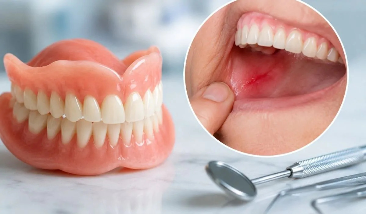
Following tooth loss, the absence of physiologic mechanical stimulation induces progressive alveolar ridge resorption. Periodontal disease, traumatic extractions, pathology, or prolonged edentulism result in severe horizontal and vertical bone deficits. This lack of structural support prevents secure placement of endosseous dental implants. To re-establish necessary bone volume and build a stable foundation, clinicians perform bone dust treatment (dental bone grafting and guided bone regeneration).
Patients considering bone augmentation often ask: "Where does this bone material come from?", "How does it integrate with living bone?", "How long does healing take?", and "Is the procedure painful?" Contemporary surgical protocols, guided bone regeneration (GBR), and bioactive barrier membranes make bone augmentation predictable and comfortable.
What Is Bone Dust Treatment?
In oral and maxillofacial surgery, the clinical definition of what is bone dust treatment is: A bone augmentation procedure in which biocompatible, osteoconductive, or osteoinductive particulate bone grafts are placed into deficient alveolar sites to stimulate new bone regeneration and establish sufficient bone width and height for implant anchorage.
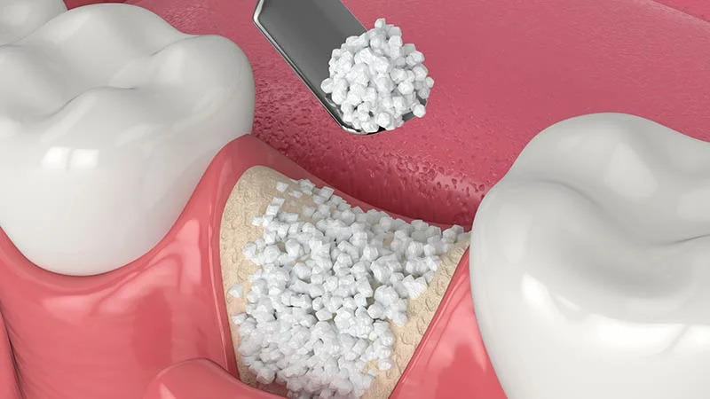
The particulate bone graft acts as a biological scaffold. Over time, host osteoblasts and vascular ingrowth colonize the matrix, gradually replacing the graft with living, vascularized host bone.
Classifications of Particulate Bone Grafts
In dental bone dust treatment, four primary graft materials are utilized based on clinical indications:
Autogenous Grafts (Autografts): Bone harvested directly from the patient (mandibular ramus or symphysis). Considered the gold standard due to its living osteogenic cellular content.
Xenografts (Bovine-Derived): Deproteinized bovine bone mineral processed into sterile, biocompatible particles. Highly effective at maintaining structural volume over long healing periods.
Allografts (Human Tissue Bank Grafts): Demineralized or mineralized freeze-dried bone allografts harvested from screened human donors and sterilized.
Alloplastic Grafts (Synthetic): Engineered biocompatible ceramics, such as beta-tricalcium phosphate (β-TCP) or hydroxyapatite.
Clinical Indications for Bone Dust Treatment for Jawbone
Performing bone dust treatment for jawbone is indicated under several anatomical conditions:
Inadequate Alveolar Ridge Dimensions: When ridge width is less than 5–6 mm or vertical bone height is under 8–10 mm.
Socket Preservation at Extraction: Placing particulate bone into extraction sockets to preserve ridge volume prior to delayed implant placement.
Maxillary Sinus Augmentation (Sinus Lift): Elevating the Schneiderian membrane and grafting particulate material to restore posterior maxillary vertical height.
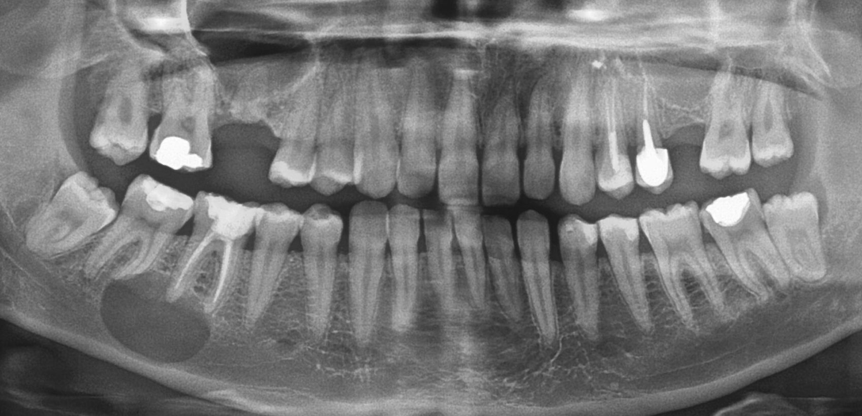
Post-Cyst Enucleation Defects: Obliterating bony cavities following cyst or tumor excision.
Periodontal Intrabony Defects: Reconstructing bone loss surrounding natural teeth to preserve periodontal stability.
How Is Bone Dust Treatment Done?
The clinical workflow explaining how is bone dust treatment done involves systematic stages:
3D Diagnostic Mapping: High-resolution Cone Beam Computed Tomography (CBCT) measures structural bone deficits in micron detail.
Local Anesthesia: The site is anesthetized; the patient experiences no pain during surgery.
Decortication and Site Preparation: A mucoperiosteal flap is elevated, and cortical bone is perforated (decortication) to promote bleeding and progenitor cell migration.
Graft Placement and Membrane Stabilization: Particulate bone is packed into the defect and covered with a resorbable collagen barrier membrane stabilized with micro-pins to prevent soft tissue ingrowth into the regenerating bone matrix.
Tension-Free Primary Closure: Mucosal flaps are released and sutured over the augmented site.
How Long Does Bone Dust Treatment Take?
Evaluating how long does bone dust treatment take involves operational time and consolidation phases:
Surgical Duration: Placing a particulate bone graft for a localized defect takes 20 to 30 minutes. Complex sinus lifts or full-arch augmentation require 45 to 60 minutes.
Bone Consolidation Window: Complete cellular remodeling and osseointegration of the graft with host bone requires 3 to 6 months before implants can support functional prosthetic loads.
Clinical Guidelines After Bone Dust Treatment
Maintaining optimal care after bone dust treatment is essential to prevent graft displacement and infection:
Avoid Spitting and Rinsing for 24 Hours: Swallow saliva to preserve the initial blood clot and stabilize graft particles.
Apply Intermittent Cold Compresses: Apply an ice pack externally to the cheek for 10-minute intervals during the first 24 hours to reduce post-operative edema.
Avoid Mechanical Pressure: Do not press the surgical site with your tongue or fingers; avoid wearing removable prostheses over the grafted zone until cleared.
Strict Smoking Cessation: Tobacco smoke compromises microvascular circulation and can cause graft failure. Abstain from smoking for at least 2 to 3 weeks.
Consume a Soft Diet: Eat lukewarm, soft, non-abrasive foods (purees, yogurt, smooth soups) during initial healing.
Comparison Matrix: Particulate Bone Graft Materials
Graft Category | Source | Osteogenic Potential | Volume Maintenance | Primary Clinical Indication |
Autograft | Patient's own bone | Superior (Contains living cells) | Moderate (Higher resorption) | Major localized bone defects |
Xenograft | Bovine bone mineral | High (Osteoconductive) | Superior (Long-term stability) | Sinus lifts, Peri-implant defects |
Allograft | Human donor tissue bank | Good (Osteoinductive) | Good | Socket preservation, Ridge defects |
Alloplastic | Synthetic bio-ceramic | Good (Osteoconductive) | Moderate | Minor contained bone defects |
Frequently Asked Questions (FAQ)
Is bone dust treatment painful during or after the procedure?
No. The procedure is performed under local anesthesia, ensuring you feel no pain during surgery. Once anesthesia wears off, mild soreness and swelling may develop, which are managed with prescribed analgesics and anti-inflammatories. Most patients resume normal daily activities within 24 to 48 hours.
Can dental implants and bone dust treatment be done at the same visit?
Yes. If baseline bone volume is sufficient to achieve initial implant stability (primary stability), simultaneous bone grafting and implant placement are routinely performed. However, if severe atrophy leaves less than 3–4 mm of bone, a staged approach is preferred: grafting is performed first, followed by implant placement 4 to 6 months later.
Can the human body reject bone dust grafts?
No. Particulate bone graft materials used in modern dentistry are approved by international health authorities (such as the FDA and CE). They undergo multi-stage purification and sterilization to remove all antigenic cellular proteins. These materials act as an inert biological scaffold and do not trigger immunological graft rejection.