Contentsexpand_more
- What is a Bony Protrusion on the Palate? (Torus Palatinus & Exostosis)
- Causes of Bony Protrusion on the Palate
- Anatomical Subtypes: Upper Palate vs. Lower Mandible
- 1. Bony Protrusion on the Upper Palate (Torus Palatinus)
- 2. Bony Protrusion on the Lower Palate / Jaw (Torus Mandibularis)
- Is a Bony Protrusion on the Palate Cancer?
- Symptoms Associated with Oral Tori
- How to Treat a Bony Protrusion on the Palate: Non-Surgical vs. Surgical
- Torus Palatinus Surgery: Clinical Excision Protocol
- Postoperative Recovery and Aftercare Guidelines
- Factors Influencing Treatment Cost
- Comparison Matrix: Torus Palatinus vs. Torus Mandibularis
- Frequently Asked Questions (FAQ)
- Can a bony protrusion on the palate grow back after surgery?
- What happens if I choose not to have the bony protrusion removed?
- Is there a risk of perforating the nasal cavity or sinus during palatal torus surgery?
Tracing your tongue along the midline of your hard palate or along the lingual floor of your mandibular arch, you may detect a stone-hard, smooth or lobulated mass projecting outward. Discovering this rigid nodule unexpectedly frequently triggers acute oncological anxiety, prompting immediate fears of a malignant bone tumor or deep-seated cystic pathology. Patients routinely evaluate: "How long has this hard bump been present?", "Is it growing?", "Is a bony protrusion on the palate dangerous?", and "Does it require surgical excision?" In maxillofacial surgery, this benign developmental exostosis is clinically recognized as a bony protrusion on the palate (termed Torus Palatinus on the maxilla or Torus Mandibularis on the mandible).
Tori are hereditary, non-neoplastic exostoses characterized by localized hyperostosis of mature, dense cortical bone. Unlike systemic bone tumors, tori are entirely benign; they lack malignant potential, do not invade adjacent neurovascular spaces, and never metastasize. However, when large tori interfere with the stability of removable prosthetics, suffer recurrent mucosal lacerations from sharp masticatory boluses, or impede normal articulation, surgical intervention is indicated. In this comprehensive clinical guide compiled by Livera Dental Clinic oral and maxillofacial surgeons, we evaluate the bony protrusion on the palate, examine anatomical variants, review triggers, and detail surgical excision protocols.
What is a Bony Protrusion on the Palate? (Torus Palatinus & Exostosis)
In oral and maxillofacial surgery, the scientific definition of a bony protrusion on the palate is: A benign, developmental, slow-growing, sessile or pedunculated exostosis of compact cortical bone projecting from the median palatine suture of the hard palate or the lingual cortex of the mandible, covered by an intact layer of normal, non-inflamed oral mucosa.
Histologically, tori do not contain internal cystic fluids or fibrous tumors; they are composed of dense, lamellar cortical bone with minimal cancellous trabeculae. Anatomical dimensions range from subtle, flat ridges to massive, multilobular masses that occupy substantial intraoral volume.
Causes of Bony Protrusion on the Palate
Diagnostic investigations identify several primary factors underlying causes of bony protrusion on the palate:
Genetic and Familial Heredity: Tori exhibit an autosomal dominant inheritance pattern with variable expressivity. Individuals with parents possessing tori have a 40% to 60% higher likelihood of developing similar exostoses.
Masticatory Hyperfunction & Bruxism: Chronic nocturnal tooth grinding and heavy bite forces transmit biomechanical shear stresses through the alveolar ridges to the median palatal suture and mandibular lingual cortex. In response, osteoblasts deposit compact bone layers to buttress mechanical loading.
Occlusal Disharmonies and Malocclusion: Unbalanced dental contacts focus functional stress across the midline of the palate.
Nutritional and Mineral Factors: Populations with marine-heavy diets high in polyunsaturated fatty acids, calcium, and vitamin D (e.g., Arctic Inuit and Mediterranean demographics) demonstrate higher epidemiological prevalences.
Age Dynamics: Tori typically become clinically apparent post-puberty, expand during the second and third decades of life, and stabilize in mature adulthood.
Anatomical Subtypes: Upper Palate vs. Lower Mandible
The anatomical site dictates clinical presentation and prosthetic implications:
1. Bony Protrusion on the Upper Palate (Torus Palatinus)
Located along the midline of the hard palate within the palatine vault.
Flat Torus: A broad-based, smooth, gently convex plateau across the palate.
Spindle Torus: An elongated, ridge-like elevation tracking the median palatal suture.
Nodular Torus: Multiple small, discrete bony protuberances coalescing along the midline.
Lobular Torus: A prominent, pedunculated mass that can descend into the oral cavity.
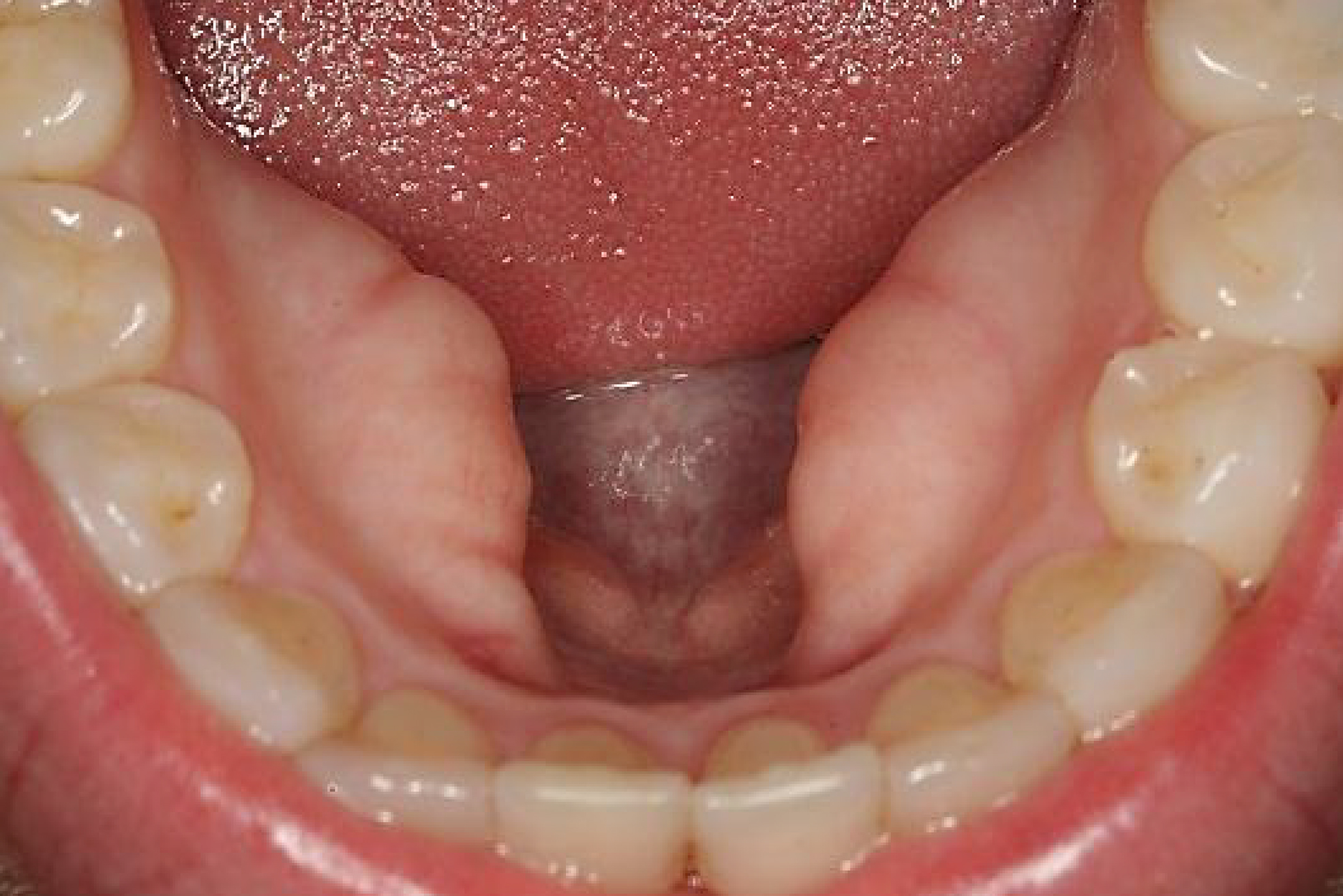
2. Bony Protrusion on the Lower Palate / Jaw (Torus Mandibularis)
Located on the lingual cortex of the mandible, superior to the mylohyoid ridge adjacent to premolars.
Presents bilaterally in over 80% of cases as smooth, symmetric nodules.
Extensive mandibular tori can displace the tongue, interfere with sublingual salivary drainage, and impede speech articulation.
Is a Bony Protrusion on the Palate Cancer?
Addressing common patient oncological concerns:
Entirely Benign: Tori are benign osteogenic formations. They carry zero malignant transformation potential and never metastasize.
Differentiation from Osteosarcomas: Malignant bone tumors expand rapidly, cause cortical perforation, induce mobility in adjacent teeth, and produce dull, continuous nocturnal bone pain. Tori develop indolently over decades and remain asymptomatic.
Clinical Vulnerability (Mucosal Ulcerations): The mucosal lining overlying a prominent torus is thin and hypovascular. Sharp foods can easily breach this epithelial envelope, resulting in persistent, painful, slow-healing ulcers.
Symptoms Associated with Oral Tori
Most individuals live with oral tori asymptomatically for decades. When symptoms manifest, they include:
Rigid, Non-Mobile Mass: A hard mass fused to the underlying palatal or mandibular bone.
Recurrent Mucosal Trauma: Painful ulcerations caused by scratch trauma from crusty bread or chips.
Phonetic Interference: Large lingual tori restricting tongue clearance during the articulation of lingual phonemes ("s", "th", "r").
Dysphagia: Sensation of food boluses catching against an enlarged palatal torus during deglutition.
Prosthetic Instability: Severe tori preclude the border seal and stable seating of removable complete or partial dentures.
How to Treat a Bony Protrusion on the Palate: Non-Surgical vs. Surgical
Evaluating clinical management options:
No Pharmacological Reversal: Tori are composed of mature cortical bone; systemic antibiotics, anti-inflammatories, and topical gels cannot resolve dense bone.
Observation as Standard Care: If a torus is small, non-ulcerated, does not interfere with speech, and no removable denture is planned, surgical excision is unnecessary.
Surgical Excision is the Definitive Treatment: When functional impairment or prosthetic fabrication demands it, surgical reduction provides permanent anatomical contouring.
Torus Palatinus Surgery: Clinical Excision Protocol
At Livera Dental Clinic, surgical removal of a palatal or mandibular torus is executed as a planned outpatient procedure:
Local Anesthesia: The palatal or lingual field is anesthetized via local infiltration, ensuring a painless operative experience.
Mucoperiosteal Flap Elevation: A linear or double-Y incision is placed along the midline of the palate, and the thin mucoperiosteum is reflected to expose the bone mass.
Piezoelectric or Rotary Osteotomy:
Using ultrasonic piezoelectric surgery or rotary surgical burs under continuous sterile saline irrigation, the bony protrusion is sectioned and shaved down to the anatomical contour of the palatal vault.
The underlying bony floor is smoothed using diamond bone files.
Flap Repositioning and Suturing: The mucosal flap is repositioned and secured with tension-free micro-sutures.
Procedure Duration: Single-site palatal reduction requires 15 to 25 minutes; complex bilateral mandibular procedures take 30 to 40 minutes.
Postoperative Recovery and Aftercare Guidelines
Following surgical torus removal:
First 24 Hours (Hemostasis): Maintain gentle pressure if a stent was delivered; avoid poking the site with your tongue. Mild pink-tinged saliva is normal.
Dietary Restrictions: Consume cool, soft, non-abrasive foods (pureed soups, yogurt, smoothies) for the first 5 to 7 days. Avoid crunchy, spicy, acidic, and piping-hot meals.
Oral Hygiene: Refrain from mechanical toothbrushing directly over the palatal incision for the first 48 hours; rinse gently with prescribed chlorhexidine gluconate.
Suture Removal: Non-resorbable sutures are removed 7 to 10 days postoperatively.
Full Mucosal Healing: Complete mucosal re-epithelialization across the palatal midline requires 2 to 3 weeks.
Factors Influencing Treatment Cost
Health regulations prohibit static surgical fees; costs depend on anatomical and procedural variables:
Dimensions and Anatomical Distribution: Excision of a small, localized midline palatal torus versus extensive bilateral mandibular tori.
Instrumentation Tier: Utilizing ultrasonic piezoelectric bone surgery units versus conventional rotary surgical handpieces.
Anesthesia Setting: Delivery under chairside local anesthesia versus IV sedation for phobic patients.
Pre-Prosthetic Surgical Packaging: Standalone reduction versus pre-prosthetic bone contouring coordinated with removable prosthetics.
Comparison Matrix: Torus Palatinus vs. Torus Mandibularis
Feature | Torus Palatinus (Upper Palate) | Torus Mandibularis (Lower Mandible) |
Anatomical Site | Midline of the hard palate | Lingual cortex adjacent to premolars |
Symmetry | Solitary midline mass | Predominantly bilateral (80%+) |
Gender Distribution | More prevalent in females | More prevalent in males |
Speech Impact | Minimal phonetic alteration | Can restrict tongue and cause lisping |
Prosthetic Interference | Breaks palatal vacuum suction seal | Impedes seating of lower partial lingual bars |
Surgical Consideration | Thin, friable overlying mucosa | Dense, thick cortical bone |
Frequently Asked Questions (FAQ)
Can a bony protrusion on the palate grow back after surgery?
Following complete surgical reduction to normal anatomical contours in adulthood, recurrence rates are minimal (approximately 1% to 2%). However, if severe nocturnal bruxism persists without occlusal nightguard therapy, minor compensatory bone remodeling can occur over decades. Wearing a nightguard helps maintain long-term results.
What happens if I choose not to have the bony protrusion removed?
If the torus does not impede mastication or speech, does not suffer recurrent ulcerations, and you do not require a removable complete or partial denture, leaving it untouched poses no medical hazard. It will not spread to other tissues or induce systemic pathology. Removal is primarily indicated for functional comfort or prosthetic accommodation.
Is there a risk of perforating the nasal cavity or sinus during palatal torus surgery?
No. Experienced oral surgeons utilize 3D cone-beam computed tomography (CBCT) to measure the vertical thickness of the palatal bone preoperatively. Resection is confined strictly to the exostotic bone projecting into the oral cavity, preserving the cortical floor of the nasal vault.