Contentsexpand_more
- What Causes a Tooth Stain?
- Classification of Tooth Stains by Chromatic Profile
- 1. Brown Tooth Stain
- 2. Black Tooth Stain
- 3. White Tooth Stain
- How to Remove Tooth Stain: Professional Clinical Protocols
- 1. Air-Flow Powder Polishing
- 2. Ultrasonic Scaling (Detertraj)
- 3. Prophylaxis and Surface Polishing
- Toothpaste for Tooth Stain and Stain Remover Products: Efficacy and Risks
- Clinical Comparison Matrix: Tooth Stain Profiles and Treatments
- Frequently Asked Questions (FAQ)
- Does professional tooth stain removal scratch or damage enamel?
- Will smoking and coffee stains return after professional cleaning?
- Can white spots on teeth be brushed away?
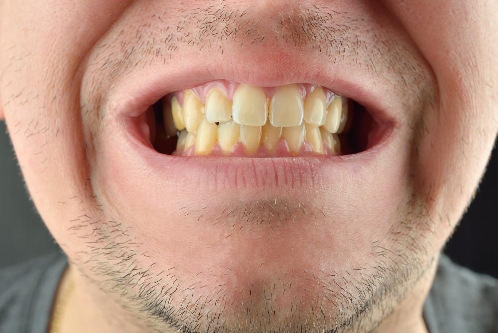
Noticing lines, speckles, or patches of discoloration across your enamel can detract from smile aesthetics and signal underlying dental health changes. A surface tooth stain creates cosmetic concern while potentially indicating microscopic enamel demineralization, calcified tartar deposits, or early carious lesions. Many individuals attempt to scrape away discolorations using aggressive home scrubbing, baking soda, or harsh chemical stain removers. However, the chromatic profile of a stain indicates its biological origin and determines the appropriate clinical removal protocol.
Black, brown, and white spots each stem from distinct physiological or pathological mechanisms within the enamel matrix. Improper DIY abrasion scratches the enamel surface, creating micro-grooves that absorb pigments faster and lead to permanent yellowing. In this comprehensive master manual compiled at Livera Dental Clinic, we evaluate what causes tooth stain, how to remove tooth stain, brown tooth stain, black tooth stain, white tooth stain, solutions for smoking tooth stain, and clinical tooth stain removal protocols.
What Causes a Tooth Stain?
When evaluating what causes tooth stain, clinical diagnostics divide etiology into extrinsic deposition and intrinsic matrix alterations:
Extrinsic Surface Staining: Caused by chromogenic pigments, dietary tannins, tobacco tar, and bacterial byproducts binding to the acquired pellicle on the outer enamel surface.
Intrinsic Matrix Discoloration: Resulting from structural changes within the enamel or underlying dentin, such as developmental fluorosis, tetracycline administration, pulpal trauma, or internal mineralization defects.
Classification of Tooth Stains by Chromatic Profile
The visual color of a tooth stain serves as a primary diagnostic indicator for selecting appropriate treatment:
1. Brown Tooth Stain
Etiology: The most prevalent stain classification. Caused by frequent intake of dark, chromogenic fluids and foods such as coffee, black tea, red wine, and dark sauces. A smoking tooth stain initially presents as dark brown bands along the cervical margins, where tar and nicotine bind to microscopic enamel imperfections.
Removal Protocol: Effectively cleared using Air-Flow high-pressure powder polishing and professional prophylaxis.
2. Black Tooth Stain
Etiology: Typically manifesting as thin lines along the gingival margin, a black tooth stain is formed by ferric sulfide precipitates produced by chromogenic anaerobic bacteria interacting with salivary iron. It can occur even in mouths with meticulous hygiene. Advanced arrested caries can also present as dark black spots.
Removal Protocol: Cleared chairside using ultrasonic scaling instruments followed by specialized polishing pastes.
3. White Tooth Stain
Etiology: Unlike dark surface deposits, a white tooth stain represents localized enamel demineralization (hypomineralization) rather than extrinsic buildup. It serves as the earliest clinical sign of incipient caries (white spot lesion) or stems from developmental fluorosis or post-orthodontic decalcification around bracket margins.
Removal Protocol: Cannot be brushed away. Treated via remineralization therapies (fluoride varnishes) or Icon Micro-Invasive Resin Infiltration to fill porous enamel structures.
How to Remove Tooth Stain: Professional Clinical Protocols
Understanding how to remove tooth stain and choosing the right tooth stain removal method requires clinical supervision. DIY abrasives scratch enamel, facilitating faster re-staining over time.
1. Air-Flow Powder Polishing
The gold standard for removing smoking, tea, and coffee deposits. Pressurized air, water, and micronized glycine or sodium bicarbonate powders are sprayed onto enamel surfaces, clearing interdental micro-grooves without damaging tooth structure.
2. Ultrasonic Scaling (Detertraj)
Indicated when surface stains are embedded within calcified calculus deposits. High-frequency ultrasonic vibrations dislodge hard tartar and black bacterial sheets.
3. Prophylaxis and Surface Polishing
Following stain removal, enamel surfaces are smoothed using rubber cups and fine-grit prophylaxis pastes. A polished, smooth enamel surface inhibits future pigment adhesion.
Toothpaste for Tooth Stain and Stain Remover Products: Efficacy and Risks
Selecting a toothpaste for tooth stain or using over-the-counter tooth stain remover devices requires caution:
Mechanism: Whitening toothpastes contain high Relative Dentin Abrasivity (RDA) particles designed to scrub away surface pigments mechanically.
Clinical Risks: Daily, long-term use of high-RDA pastes abrades enamel, exposing the underlying yellow dentin and increasing surface roughness.
Recommended Usage: Abrasive stain toothpastes should be limited to once or twice weekly, using low-abrasivity fluoride formulations for daily hygiene.
Clinical Comparison Matrix: Tooth Stain Profiles and Treatments
Stain Profile / Color | Primary Etiology | DIY Removal Feasibility | Primary Clinical Treatment |
Brown Stain | Coffee, tea, tobacco tar accumulation | Very Low (Fresh stains only) | Air-Flow Polishing + Prophylaxis |
Black Stain | Chromogenic bacterial deposits / Tartar | Impossible | Ultrasonic Scaling + Air-Flow |
White Stain | Enamel demineralization (White spot) | IMPOSSIBLE | Icon Resin Infiltration / Fluoride |
Yellow Stain | Surface bio-film / Enamel thinning | Partial (Daily brushing) | Scaling + In-Office Bleaching |
Frequently Asked Questions (FAQ)
Does professional tooth stain removal scratch or damage enamel?
No, professional stain removal performed in a clinical setting (such as Air-Flow and ultrasonic scaling) does not scratch or weaken enamel. Air-Flow micro-powders and prophylaxis pastes are softer than calcified enamel matrix, removing extrinsic deposits safely.
Will smoking and coffee stains return after professional cleaning?
Yes, continuous exposure to tobacco, dark teas, and coffee will gradually re-deposit pigments on enamel over time. However, professional prophylaxis polishes the enamel surface, making it smoother and delaying re-staining. Maintaining brushing twice daily, using interdental brushes, and scheduling routine cleanings every 6 months minimizes stain recurrence.
Can white spots on teeth be brushed away?
No, white spot stains cannot be brushed away. White spots represent areas where enamel has lost calcium and phosphate minerals, becoming porous (incipient decay or decalcification). Brushing cannot restore lost mineral structure. Treatment requires clinical remineralization or Icon Micro-Invasive Resin Infiltration to fill the enamel pores with fluid resin.