Contentsexpand_more
- What is Peri-Implantitis?
- What Causes Peri-Implantitis?
- Peri-Implantitis Symptoms and Diagnostic Markers
- Peri-Implant Mucositis vs. Peri-Implantitis
- Peri-Implantitis Treatment: Clinical Decontamination Protocols
- 1. Non-Surgical Mechanical and Laser Decontamination (Early Defect)
- 2. Open-Flap Surgical Debridement and Implantoplasty
- 3. Regenerative Bone Surgery (Grafts and Barrier Membranes)
- 4. Explantation (Implant Removal as Last Resort)
- Factors Influencing Peri-Implantitis Treatment Cost
- Comparison Matrix: Peri-Implant Mucositis vs. Peri-Implantitis
- Frequently Asked Questions (FAQ)
- Can a mobile dental implant be saved with peri-implantitis treatment?
- Will antibiotics alone cure peri-implantitis?
- How can I prevent bone loss around my dental implants?
Years after having a dental implant surgically placed into your jawbone and enjoying the comfort of chewing as with a natural tooth, you may suddenly observe bleeding during toothbrushing, marginal erythema, or dull localized tenderness around the restoration. Many patients dismiss these symptoms, believing the misconception that "titanium implants are artificial and immune to disease." While titanium cannot decay, the surrounding alveolar bone and peri-implant mucosa remain susceptible to destructive microbial attacks. Progressive inflammatory bone resorption surrounding an osseointegrated fixture is clinically recognized in periodontology as peri-implantitis.
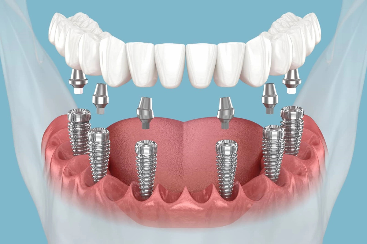
Natural teeth are suspended within the alveolar housing via the periodontal ligament, a vascular, cell-rich shock-absorbing apparatus with an organized supracrestal connective tissue attachment. In contrast, dental implants achieve direct bone-to-implant contact (osseointegration) without a true periodontal ligament barrier. Because peri-implant tissues exhibit reduced vascularity and parallel-running collagen fibers that fail to insert into the titanium collar, microbial biofilm penetrating subgingivally triggers rapid, circumferential bone resorption. Without timely intervention, terminal loss of osseointegration occurs, resulting in implant failure.
What is Peri-Implantitis?
In clinical periodontology and oral implantology, the medical definition of what is peri-implantitis is: A plaque-associated pathological condition occurring in tissues around dental implants, characterized by inflammation in the peri-implant mucosa and progressive loss of supporting alveolar bone beyond physiological remodeling.
Peri-implant diseases are categorized into two progressive stages:
Peri-Implant Mucositis: An inflammatory lesion strictly confined to the peri-implant soft tissues without loss of supporting alveolar bone. Analogous to gingivitis, it is fully reversible with non-surgical biofilm debridement.
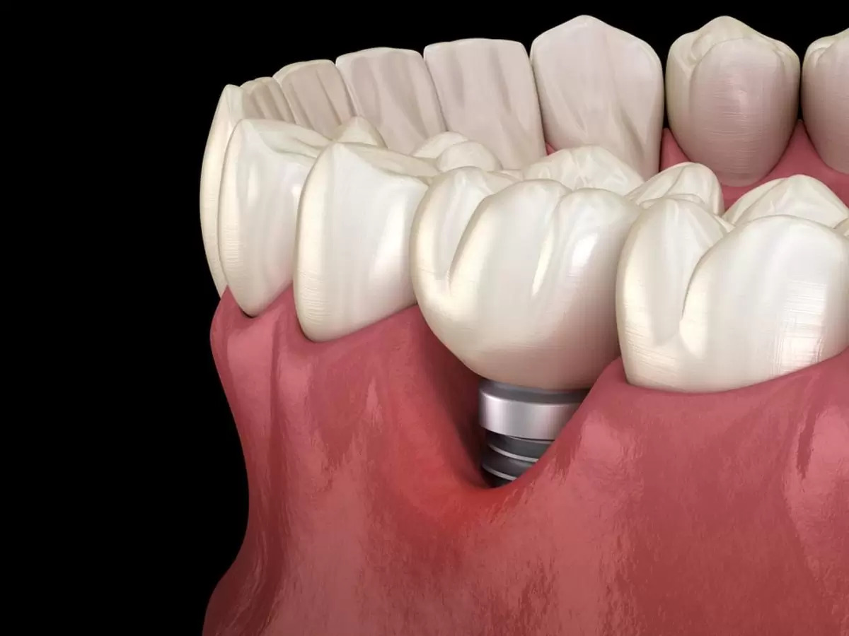
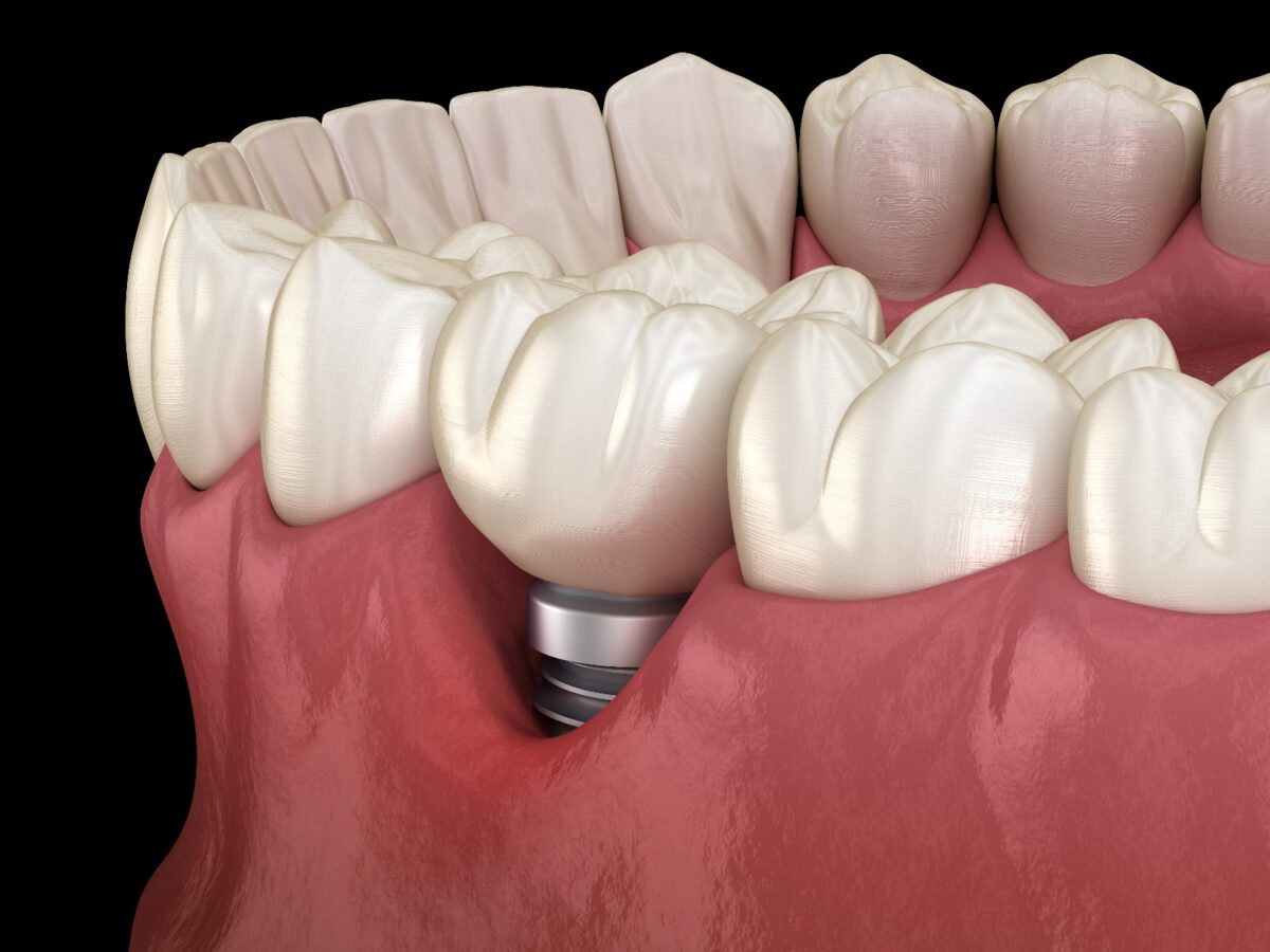
Peri-Implantitis: The unmitigated progression of mucosal inflammation into the underlying osseous bed, resulting in deepening pathological pockets, suppuration, and irreversible circumferential bone cratering around titanium threads.
What Causes Peri-Implantitis?
Clinical investigations identify several primary factors underlying the etiology of peri-implantitis:
Inadequate Oral Hygiene & Plaque Biofilm Accumulation: Failure to decontaminate prosthetic superstructures using interdental proxy brushes, specialized implant floss, or oral irrigators permits mature anaerobic biofilms (Porphyromonas gingivalis, Treponema denticola) to colonize the titanium interface.
History of Periodontitis: Patients who lost natural dentition to severe periodontal disease harbor persistent periodontal pathogens that colonize peri-implant crevices.
Tobacco Smoking: Nicotine induces peripheral vasoconstriction, impairs local leukocyte phagocytosis, downregulates osteoblastic activity, and elevates bone resorption rates by three- to four-fold.
Retained Subgingival Excess Cement: Extruded, polymerized resin or zinc cement remnants left behind during fixed crown cementation provoke severe foreign-body reactions and rapid osteoclastogenesis.
Occlusal Overload and Biomechanical Stress: Parafunctional bruxism and non-axial cantilever forces generate micro-strains at the bone-implant interface, accelerating bone loss in the presence of inflammation.
Uncontrolled Systemic Conditions: Poorly controlled Type 2 diabetes mellitus, systemic osteoporosis, and immunosuppressive medications compromise tissue regeneration.
Peri-Implantitis Symptoms and Diagnostic Markers
Because bone loss is largely painless in its early stages, identifying clinical peri-implantitis symptoms is critical:
Profuse Bleeding on Probing (BOP): Bleeding triggered upon gentle probing or during daily mechanical toothbrushing.
Mucosal Erythema and Edema: The peri-implant collar appears swollen, boggy, erythematous, or deep purple.
Suppuration and Purulent Exudate: Milking the peri-implant margin yields purulent, foul-tasting discharge from the pocket.
Gingival Recession & Exposed Titanium Threads: Progressive crestal resorption exposes rough dark grey implant threads above the soft-tissue line.
Halitosis and Metallic Dysgeusia: Persistent foul breath generated by volatile sulfur compounds produced by trapped anaerobic bacteria.
Implant Mobility (Terminal Sign): A mobile fixture indicates total loss of osseointegration, representing end-stage implant failure.
Peri-Implant Mucositis vs. Peri-Implantitis
Differentiating between these diagnostic categories establishes the treatment plan:
Tissue Involvement: Mucositis is confined strictly to the soft mucosal envelope; bone remains intact. Peri-implantitis involves soft tissue inflammation paired with progressive bone loss.
Radiographic Presentation: Intraoral radiographs of mucositis show stable crestal bone levels identical to baseline post-placement images. Peri-implantitis displays characteristic saucer-shaped or crater-like radiolucencies around fixture threads.
Reversibility: Mucositis is completely reversible with professional prophylaxis and home-care optimization within 7 to 14 days. Peri-implantitis involves structural bone deficits that require surgical decontamination and regenerative therapies.
Peri-Implantitis Treatment: Clinical Decontamination Protocols
At Livera Dental Clinic, peri-implantitis treatment is executed through phased protocols based on bone defect morphology:
1. Non-Surgical Mechanical and Laser Decontamination (Early Defect)
Indicated when crestal bone loss is minimal (under 2 mm):
Mechanical Debridement: Calculus and biofilms are cleared using non-abrasive titanium, carbon-fiber, or polyetheretherketone (PEEK) ultrasonic tips.
Laser-Assisted Sterilization (Er:YAG / Diode Lasers): Laser energy penetrates microscopic titanium surface porosities, eradicating up to 99% of bacterial biofilms without overheating surrounding bone.
Local Chemotherapeutics: Subgingival delivery of minocycline microspheres or chlorhexidine chips suppresses remaining pathogens.
2. Open-Flap Surgical Debridement and Implantoplasty
When bone loss is moderate, a full-thickness mucoperiosteal flap is elevated for direct access:
Granulation tissue is meticulously curetted from the defect.
Implantoplasty: The exposed, rough titanium threads are mechanically ground down, smoothed, and polished to a mirror finish using fine diamond and silicone burs. This eliminates microscopic niches for plaque retention.
3. Regenerative Bone Surgery (Grafts and Barrier Membranes)
When circumferential crater defects with intact bony walls are present, regenerating lost bone is pursued:
Following chemical and laser decontamination, the bony crater is packed with particulate bone allografts/xenografts and covered with a resorbable collagen membrane.
Platelet-rich fibrin (PRF) concentrates are integrated to deliver growth factors that promote re-osseointegration before tension-free primary closure.
4. Explantation (Implant Removal as Last Resort)
When bone loss exceeds 50% of the fixture length, mobility is present, or the implant position is prosthetically unrestorable, the fixture is atraumatically retrieved using reverse-torque explantation kits. The socket is debrided and grafted for future replacement.
Factors Influencing Peri-Implantitis Treatment Cost
Health regulations restrict stating uniform treatment fees; peri-implantitis treatment costs depend on case complexity. Primary variables include:
Severity of Bone Resorption & Procedure Class: Non-surgical laser-assisted debridement versus complex open-flap regenerative bone grafting.
Volume of Biomaterial Bone Grafts and Membranes: The cubic centimeters (cc) of particulate bone graft materials, barrier membranes, and PRF preparations required.
Advanced Technology Employed: Utilization of Er:YAG hard-tissue lasers, specialized titanium ultrasonic instruments, and glycine air-polishing units.
Prosthetic Modification or Replacement: Need to detach, modify, contour, or completely remake the overlying crown to eliminate cement traps and establish cleansable margins.
Comparison Matrix: Peri-Implant Mucositis vs. Peri-Implantitis
Clinical Criteria | Peri-Implant Mucositis | Peri-Implantitis |
Anatomical Boundary | Soft mucosal tissue only | Soft tissue and alveolar bone |
Marginal Bone Loss | Zero (Crestal bone intact) | Progressive (Visible around threads) |
Radiographic Defect | None | Saucer-shaped cratering |
Probing Pocket Depth | Mild pseudopockets (under 4 mm) | Pathological deep pockets (5 mm or greater) |
Spontaneous Suppuration | Rare | Common diagnostic hallmark |
Therapeutic Reversibility | 100% reversible | Non-reversible without surgery |
Primary Clinical Protocol | Prophylaxis, air-polishing, hygiene | Laser debridement, bone graft, implantoplasty |
Frequently Asked Questions (FAQ)
Can a mobile dental implant be saved with peri-implantitis treatment?
No. Once a dental implant exhibits clinical mobility, it indicates that osseointegration has been lost. Unlike natural teeth with periodontal ligaments that can tighten after therapy, an unanchored titanium fixture cannot biologically reattach to bone once mobile. The mobile implant must be removed, the site grafted with bone substitutes, and a new implant planned after 4 to 6 months of healing.
Will antibiotics alone cure peri-implantitis?
No. Systemic antibiotics diffuse through blood vessels into local tissues, but they cannot penetrate the bacterial biofilm adhering to the physical titanium surface. Antibiotics may temporarily diminish swelling and acute symptoms, but the bacterial reservoir remains on the threads. Mechanical removal, chemical decontamination, and surgical correction are necessary to resolve the infection.
How can I prevent bone loss around my dental implants?
Preventing peri-implant disease relies on meticulous daily hygiene and scheduled recall appointments. Use interdental proxy brushes with plastic-coated wires, spongy implant floss, and water flossers daily. Visit your dental hygienist every 4 to 6 months for professional maintenance and annual periapical radiographs to monitor crestal bone levels. Smoking cessation significantly lowers long-term risk.