Contentsexpand_more
- What is Peri-implantitis?
- What Causes Peri-implantitis?
- Symptoms of a Failing Dental Implant
- How to Treat Implant Infection?
- 1. Non-Surgical Debridement and Decontamination
- 2. Advanced Laser Peri-implantitis Therapy
- 3. Surgical Regenerative Therapy
- How Long Does Peri-implantitis Treatment Take?
- Frequently Asked Questions (FAQ)
- Does an implant diagnosed with active peri-implantitis always require extraction?
- Will I experience sharp pain during or after implant infection treatment?
- What home-care steps are mandatory to protect dental implants from infection?
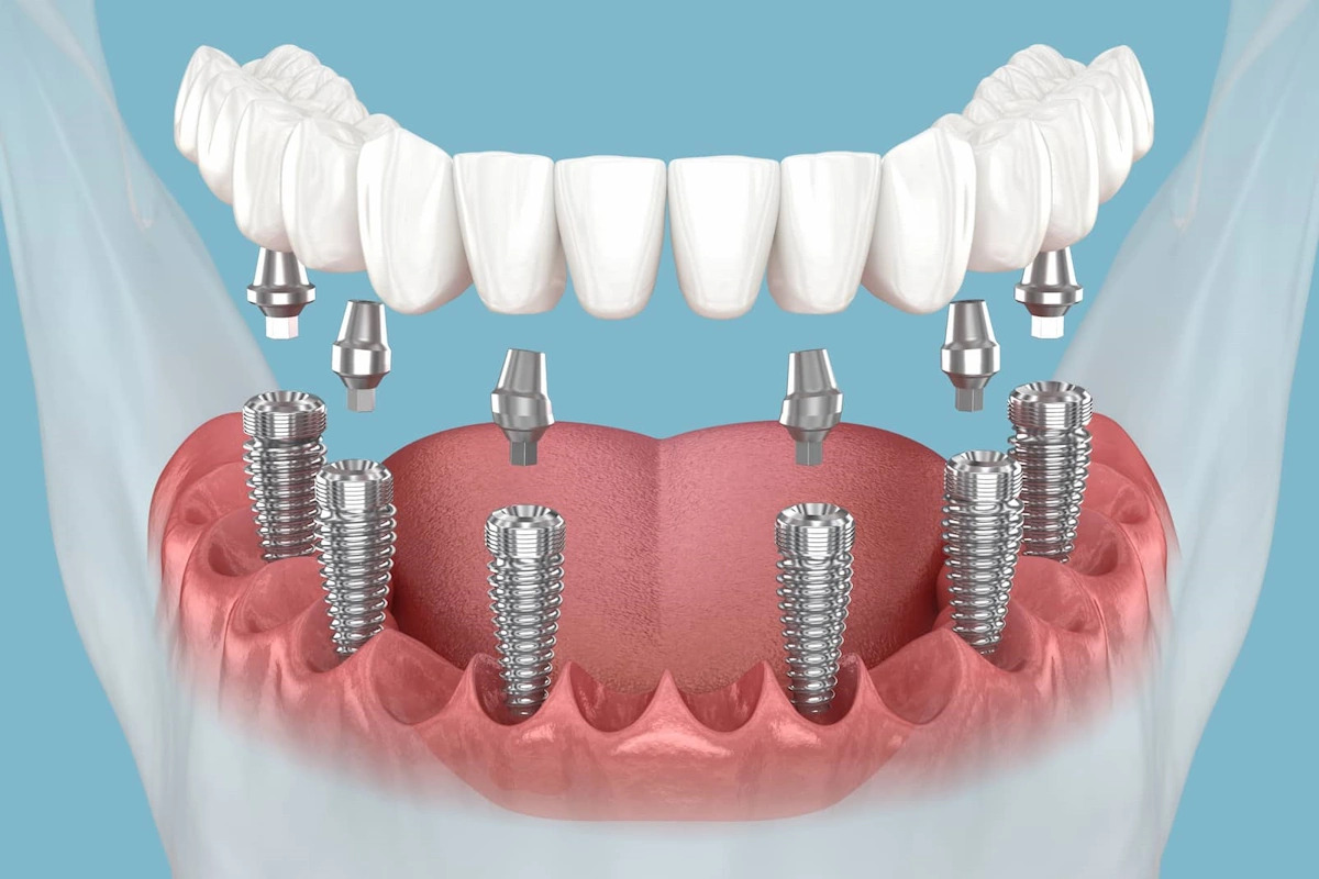
Within contemporary restorative dentistry, dental implants represent the most successful and biomechanically sound modality to replace missing teeth. Engineered from medical-grade titanium or biocompatible zirconium, these artificial roots are structured to forge a seamless biological bond (osseointegration) with the surrounding jawbone, designed to achieve lifetime functional durability. However, as extensively emphasized across global medical literature, implants require identical, if not more meticulous, protective vigilance against aggressive bacterial infections than natural dentition. Just as chronic periodontitis degrades the supporting alveolar bone of a natural tooth leading to mobility, pathogenic colonization around an implant can rapidly induce structural degradation and subsequent fixture loss (failing dental implant / implant failure).
Characterized by the progressive destruction of the surrounding soft mucosal tissues and, most critically, the underlying alveolar bone housing the structural fixture, this pathology stands as a highly complex issue in contemporary implantology. Peri-implantitis treatment can be successfully executed. When caught during early to moderate stages, it is fully viable to salvage the implant fixture and halt progressive bone loss entirely. This comprehensive manual evaluates what causes peri-implantitis, its progressive clinical symptoms, and advanced chairside eradication techniques.
What is Peri-implantitis?
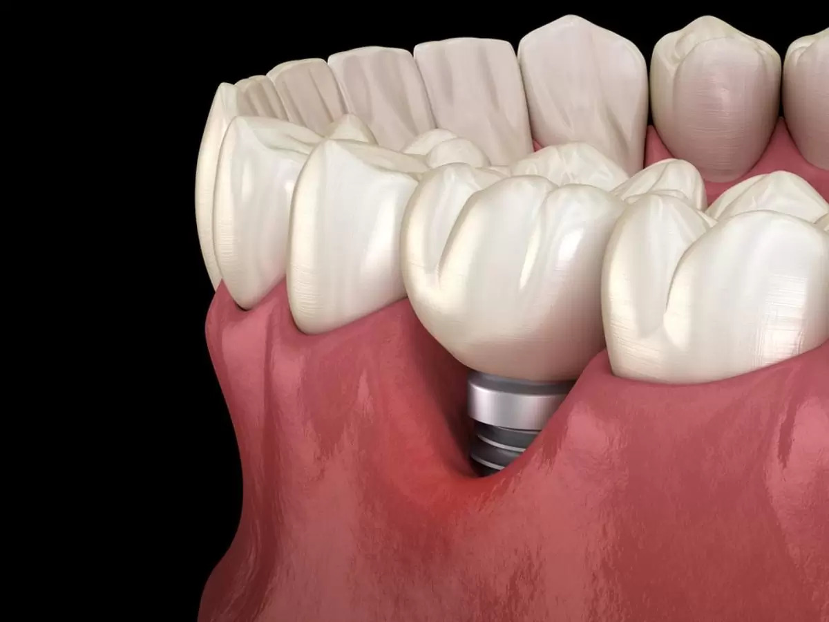
The primary baseline query raised by patients navigating localized discomfort or structural discharge around their existing fixtures is what is peri-implantitis. Peri-implantitis is defined as a destructive, progressive inflammatory condition targeted to the hard osseous (bone) and soft mucosal tissues surrounding an osseointegrated dental implant, triggered entirely by virulent anaerobic bacterial biofilms.
Clinically mapping this disease requires a clear distinction across two phenotypes: if the active inflammatory response is localized strictly within the surrounding peri-implant mucosa without compromising bone parameters, it is classified as peri-implant mucositis (the structural equivalent of gingivitis, which is fully reversible upon professional scaling). However, if the bacterial lesion expands deeper, breaching the mucosal boundary to cause direct resorption (melting) of the underlying alveolar jawbone supporting the fixture, the pathology progresses into active peri-implantitis.
What Causes Peri-implantitis?
The long-term infection and failure of an established dental implant rarely stem from an isolated mechanical anomaly. Exploring what causes peri-implantitis highlights several primary multi-factorial vectors:
Deficient Oral Hygiene and Virulent Biofilms: This represents the primary causative catalyst. Although dental implants cannot develop dental caries, their surrounding soft tissue seals lack the complex ligamentous defense of natural teeth, making them highly vulnerable to stagnant bacterial plaque. Neglecting specialized interdental cleaning beneath implant-supported prosthetics allows subgingival biofilms to proliferate rapidly.
Heavy Tobacco and Smoking Habits: Tobacco smoke drives severe, chronic vasoconstriction across the peri-implant capillary networks, actively starving the local tissues of oxygenated blood flow and neutralizing maternal immune cell migration. Statistical metrics show that smokers present a 4-to-5-fold higher risk of developing peri-implant bone loss.
Uncontrolled Systemic Pathologies: Unregulated Type-2 Diabetes Mellitus and progressive Osteoporosis severely compromise local osseous metabolism, decreasing bone density and lowering tissue resistance against microbial attacks.
Prosthetic Design Flaws and Retained Cement Scraps: Excess submucosal luting cement (glue) forced beneath the gingival margin during the fixation of implant crowns acts as an aggressive foreign-body irritant, inducing rapid, localized bone resorption. Additionally, non-cleansable prosthetic contours prevent effective mechanical plaque control.
Excessive Biomechanical Overload (Bruxism): Intense, unmitigated nocturnal teeth grinding directs destructive vertical and lateral vectors into the fixture, triggering microscopic fractures at the bone-to-implant interface that welcome bacterial infiltration.
Symptoms of a Failing Dental Implant
Identifying the early warning signs of progressive peri-implant degradation is vital. The most distinct failing dental implant symptoms requiring immediate clinical triage include:
Continuous erythema (redness), edematous swelling, and spontaneous bleeding of the mucosa surrounding the implant neck.
Suppuration (pus discharge) emitting from the peri-implant sulcus upon applying minor digital pressure to the gums.
Persistent halitosis (bad breath) and a constant unpleasant metallic taste in the oral cavity.
Dull, throbbing, or deep localized bone pain during mastication or functional loading.
Advanced-stage macroscopic mobility of the implant fixture. If a dental implant exhibits distinct mobility, it signifies a total loss of osseous support, rendering the fixture unsalvageable and requiring absolute surgical removal.
How to Treat Implant Infection?
Once structural bone loss is radiographically verified around a fixture, immediate intervention must be initiated. So, what is peri-implantitis treatment and how to treat implant infection successfully within our specialized surgical wing? The clinical workflow is structured across non-surgical and advanced micro-surgical phases:
1. Non-Surgical Debridement and Decontamination
The initial phase focuses on completely removing hard calculus and subgingival biofilm layers. To prevent scratching or altering the delicate micro-textured titanium surface of the implant, traditional stainless steel scalers are strictly banned; instead, advanced carbon-fiber, plastic, or pure titanium curettes are leveraged alongside localized antimicrobial irrigants and sustained-release antibiotic gels.
2. Advanced Laser Peri-implantitis Therapy
This represents the cutting-edge innovation yielding premium clinical success at Livera Dental Clinic. Thoroughly cleansing bacterial biofilms from the deep, complex micro-grooves of a threaded titanium implant using manual hand instruments is mechanically impossible. Utilizing specific wavelengths during laser peri-implantitis therapy allows our specialists to completely vaporize the subgingival bacterial biofilm at a cellular level without causing thermal alterations to the titanium core. The laser energy sanitizes the absolute deepest limits of the fixture threads.
3. Surgical Regenerative Therapy
In presentations with advanced horizontal or vertical bone defects, the soft tissue is surgically reflected (flap surgery) to achieve direct visual access. Following exhaustive chemical and laser-driven surface decontamination of the exposed implant threads, specialized bone grafts (bone powders) combined with barrier membranes are adapted into the osseous defect. The site is closed with microsurgical sutures, aggressively stimulating guided bone regeneration (GBR) to rebuild the lost alveolar housing around the fixture.
How Long Does Peri-implantitis Treatment Take?
The active clinical sessions associated with direct decontamination can require 1 to 3 targeted appointments. However, if micro-surgical bone grafting is performed to reconstruct the lost housing, the biological timeline required for the bone powders to fully integrate with the patient's native skeleton and mature into dense bone spans 3 to 6 months. Throughout this phase, the patient must maintain immaculate hygiene metrics and attend routine follow-up observations (typically once a month) to verify clinical healing.
Frequently Asked Questions (FAQ)
Does an implant diagnosed with active peri-implantitis always require extraction?
No, an implant diagnosed with active peri-implantitis does not automatically require surgical extraction. If the inflammatory process is intercepted during early or moderate stages, where bone loss has not surpassed 50% of the implant length, and the fixture remains firmly locked within the bone (zero mobility), advanced laser decontamination and bone grafting can successfully salvage the implant in 80-90% of cases. However, if true mobility is recorded, the osseointegration is lost, making extraction mandatory.
Will I experience sharp pain during or after implant infection treatment?
Absolute zero pain is experienced during the active treatment procedures. All debridement, micro-surgical tissue reflection, and laser therapies are executed under profound local anesthesia, fully desensitizing the working quadrant. Following advanced surgical bone reconstruction, mild localized swelling and dull tenderness are expected for 48-72 hours, which are seamlessly controlled using prescribed anti-inflammatory and analgesic pharmaceuticals.
What home-care steps are mandatory to protect dental implants from infection?
The long-term structural survival of a dental implant is heavily dependent on daily customized home hygiene. Bypassing traditional flossing, patients must utilize thick-ended specialized floss (superfloss) to sweep beneath prosthetic margins, coupled with high-efficiency hydro-magnetic oral irrigators (water flossers) to flush out subgingival debris from the implant threads daily. Attending professional periodontist hygiene reviews at least twice a year accompanied by baseline digital radiographs is equally essential.