Contentsexpand_more
- What is Abfraction?
- Causes of Abfraction
- Abfraction vs. Abrasion: Clinical Differences
- Symptoms of Abfraction Lesions
- Abfraction Treatment: Clinical Modalities
- 1. Occlusal Equilibration and Bite Balancing
- 2. Custom Hard Occlusal Splints (Nightguards)
- 3. Biomimetic Flexible Resin Restorations (Cervical Bonding)
- 4. Periodontal Mucogingival Surgery (Connective Tissue Grafts)
- Factors Influencing Abfraction Treatment Cost
- Comparison Matrix: Abfraction vs. Abrasion vs. Erosion
- Frequently Asked Questions (FAQ)
- Why do fillings placed in my cervical notches keep falling out?
- Can an untreated abfraction cause my tooth to break off?
- Will my abfraction stop progressing if I stop brushing hard?
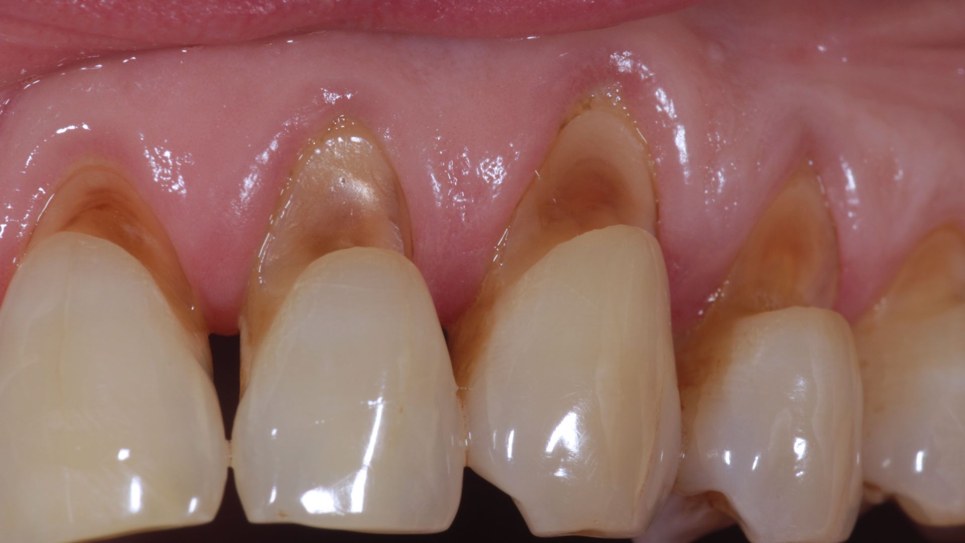
Running your fingernail along the junction where your tooth meets the gumline, you may detect a distinct, sharp V-shaped wedge, notch, or ledge. Inhaling cool air or taking a sip of chilled water often triggers a sharp, localized zing. Many patients assume these cervical defects stem from aggressive scrub toothbrushing or cervical dental caries. In clinical restorative dentistry, a wedge-shaped cervical defect driven by biomechanical tooth flexure under occlusal loading is recognized as abfraction.
Human teeth act like load-bearing architectural columns; under heavy masticatory forces, they flex and bend at a microscopic level. The fulcrum of this flexural movement is the cementoenamel junction (CEJ) at the cervical neck, where enamel is naturally thin. Excessive occlusal forces concentrate tensile stress at this cervical margin, causing brittle hydroxyapatite prisms to micro-fracture.
What is Abfraction?
In restorative dentistry, the clinical definition of what is abfraction is: A non-carious cervical tooth defect (NCCL) characterized by a wedge-shaped loss of hard tissue with sharp internal line angles at the cementoenamel junction, resulting from biomechanical loading forces that cause tooth flexure and tensile stress concentration.
Enamel resists compressive forces well but has low tensile strength. When non-axial lateral vectors flex the clinical crown during mastication or clenching, the cervical margin experiences alternating tensile and compressive stresses, causing enamel rods to disrupt and dislodge.
Causes of Abfraction
Clinical diagnostic investigations identify several primary factors underlying the causes of abfraction:
Nocturnal Bruxism and Clenching: The leading global trigger. Involuntary nighttime jaw clenching exerts heavy shear forces across the occlusal table; repeated lateral excursions flex the tooth crown and fatigue cervical enamel.
Occlusal Premature Contacts and Interferences: A single cuspal high spot encountering premature contact during mandibular excursion introduces heavy cantilever loads during chewing.
Crossbites and Malocclusions: Tipped, rotated, or lingually locked teeth receive non-axial vectors, compounding cervical tensile stress concentrations.
Posterior Bite Collapse (Loss of Molar Support): Missing posterior molars transfer chewing loads anteriorly onto premolars and canines, which can accelerate cervical structural breakdown.
Compromised Periodontal Bone Support: When bone levels recede from periodontitis, the tooth's center of resistance shifts apically, increasing the bending moment on the cervical region.
Abfraction vs. Abrasion: Clinical Differences
Understanding the difference between abfraction and abrasion is guided by distinct diagnostic criteria:
Etiological Mechanism: Abrasion is caused by external friction from hard toothbrush bristles, high-RDA abrasive whitening pastes, or foreign objects. Abfraction is driven by internal tooth flexure under occlusal force.
Morphological Geometry: Abrasion lesions present as broad, shallow, dish-shaped U-shaped defects with rounded, smooth borders. Abfraction lesions present as narrow, deep, wedge-shaped V-notches with sharp margins.
Spatial Distribution: Abrasion tracks across facial surfaces where toothbrushing scrub pressure peaks (typically canines and premolars). Abfraction can develop on a single tooth receiving isolated occlusal trauma and may extend subgingivally.
Combined Pathology: These conditions frequently coexist. An abfraction micro-fracture can be exacerbated by abrasive toothbrushing and acidic dietary erosion, accelerating defect progression.
Symptoms of Abfraction Lesions
Typical clinical presentations associated with abfraction lesions include:
Palpable V-Shaped Cervical Notch: A sharp ledge at the gumline that catches fingernails or dental probes.
Acute Cervical Hypersensitivity: Exposed dentinal tubules trigger sharp, fleeting pain when exposed to cold fluids, sweet foods, or cold air.
Food Impaction and Floss Catching: Fibrous food debris packing into the cervical notch, causing localized gingival inflammation.
Recurrent Restoration Debonding: Fillings placed into active abfraction defects dislodge repeatedly as continued tooth flexure breaks the adhesive bond.
Structural Weakness: Severe notches can hollow out the cervical neck, increasing the risk of coronal tooth fracture under bite pressure.
Abfraction Treatment: Clinical Modalities
At Livera Dental Clinic, abfraction treatment pairs restorative rebuilding with management of the underlying occlusal forces:
1. Occlusal Equilibration and Bite Balancing
The foundational step involves using digital occlusion sensors or articulating paper to locate premature contacts and interferences. Micro-adjustments neutralize lateral shear vectors on the affected tooth.
2. Custom Hard Occlusal Splints (Nightguards)
For patients with nocturnal bruxism, a custom 3D digital hard-acrylic occlusal splint shields restorations and disperses clenching forces evenly across the arches.
3. Biomimetic Flexible Resin Restorations (Cervical Bonding)
Restoring deep V-shaped notches requires materials that flex with the tooth rather than brittle micro-hybrids:
Flowable Composites with Low Modulus of Elasticity: Flexible micro-hybrid resins bend with tooth movement, resisting marginal debonding.
Resin-Modified Glass Ionomer (RMGI) Cements: Chemically bond to dentin and cementum, release fluoride, and flex with the tooth structure under functional load.
4. Periodontal Mucogingival Surgery (Connective Tissue Grafts)
When deep abfraction defects are accompanied by severe marginal gingival recession, restorative contouring is followed by a subepithelial connective tissue graft to restore pink aesthetics and cover exposed roots.
Factors Influencing Abfraction Treatment Cost
Regulatory protocols prohibit static pricing; abfraction treatment costs depend on case complexity. Primary variables include:
Number and Depth of Cervical Lesions: Treating a localized notch on a single tooth versus multiple abfraction defects across an entire arch.
Selected Biomaterial Class: Low-modulus flowable composite resins, bioactive glass-ionomers, or self-etching adhesives.
Adjunctive Occlusal Therapy and Splints: Incorporating occlusal equilibration analysis and custom 3D digital hard nightguard fabrication.
Need for Periodontal Surgery: Adding connective tissue grafting to cover associated gingival recession.
Comparison Matrix: Abfraction vs. Abrasion vs. Erosion
Criterion | Abfraction | Abrasion | Erosion |
Primary Driver | Occlusal stress and tooth flexure | Mechanical scrub friction | Chemical acid dissolution |
Lesion Geometry | Deep, angular V-shaped notch | Shallow, smooth U-shaped dish | Rounded, cupped, glossy depression |
Caries Involvement | Non-carious hard tissue loss | Non-carious | Non-carious |
Common Location | Cervical neck (Can affect single teeth) | Facial margins along brush paths | Lingual surfaces, occlusal pits |
Primary Therapy | Occlusal adjustment + Splint + Flex-filling | Soft brush + Low-RDA paste + Bonding | Dietary control + Remineralization |
Frequently Asked Questions (FAQ)
Why do fillings placed in my cervical notches keep falling out?
Cervical restorations frequently debond because the underlying tooth flexure has not been addressed. Every time you bite or clench, the tooth flexes at the cervical margin. If a rigid, high-modulus composite is placed without adjusting the bite, the tooth's flexure breaks the adhesive bond. Successful care requires balancing occlusal contacts, placing flexible low-modulus restorative materials, and using a nightguard.
Can an untreated abfraction cause my tooth to break off?
Yes. Abfraction is a progressive defect. As occlusal forces continue unmitigated, the V-shaped notch deepens toward the pulp chamber. The coronal-radicular junction thins significantly, leaving the tooth vulnerable to horizontal fracture at the gumline under normal chewing loads.
Will my abfraction stop progressing if I stop brushing hard?
No. Switching to a soft toothbrush reduces mechanical abrasion, but it does not stop abfraction. Because abfraction is driven by occlusal forces and bruxism, clenching will continue to flex the tooth and dislodge cervical enamel rods even without toothbrush friction. A clinical bite evaluation is necessary to manage the root cause.