Contentsexpand_more
- What is Gum Peeling?
- What Causes Dark Gums and Melanin Pigmentation?
- How is Gum Peeling Done?
- Gum Peeling Recovery: Healing Timeline and Stages
- Postoperative Care Guidelines Following Gum Peeling
- Factors Influencing Gum Peeling Cost
- Comparison Matrix: Laser Gum Peeling vs. Conventional Surgical Abrasion
- Frequently Asked Questions (FAQ)
- Does gum peeling hurt during or after the procedure?
- Can dark spots return after gum peeling?
- Does gum peeling damage tooth enamel or underlying roots?
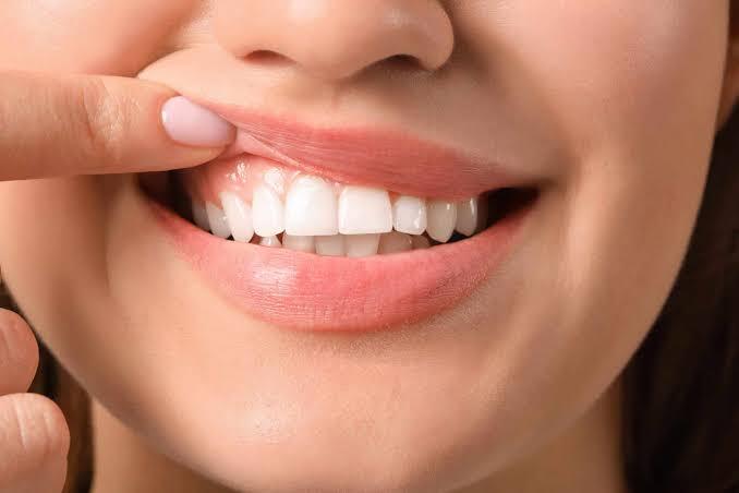
When evaluating your smile in the mirror, the alignment, shade, and contour of your dental crowns may appear flawless; however, dark brown, purple, or black patches across your gingival margins can detract from your overall aesthetics. Many patients assume this discoloration is an irreversible consequence of tobacco use, poor circulation, or a genetic condition that must be tolerated. In contemporary aesthetic periodontology, gently resurfacing the hyperpigmented superficial layer of the gums to reveal healthy, uniform pink tissue is recognized as gum peeling (clinically termed gingival depigmentation).
The physiological pigment responsible for gingival coloration is melanin, synthesized by melanocytes located in the basal epithelial layer. In certain individuals, genetic background, higher constitutional skin phototypes, hormonal fluctuations, or prolonged tobacco use stimulate melanocytes to deposit excess pigment. This is not a pathology; it is a physiological hyperpigmentation. The objective of cosmetic periodontal therapy is to restore a uniform coral-pink frame that complements your smile.
What is Gum Peeling?
In aesthetic periodontology, the clinical definition of what is gum peeling is: A cosmetic periodontal resurfacing procedure designed to ablate, de-epithelialize, or vaporize excess melanin pigmentation located within the gingival epithelium using soft-tissue dental lasers, rotary diamond instruments, or micro-surgical scalpels, allowing non-pigmented connective tissue to regenerate into a uniform coral-pink hue.
This treatment does not involve cutting away functional gum margins or reducing tissue volume; it is a superficial de-epithelialization protocol targeting the stratum basale.
What Causes Dark Gums and Melanin Pigmentation?
Diagnostic evaluations identify several primary factors underlying dark gingival pigmentation:
Genetic and Racial Predisposition: Individuals of Mediterranean, African, Asian, or Hispanic heritage naturally possess active melanocytes that produce higher baseline melanin.
Tobacco Smoking (Smoker's Melanosis): Nicotine and thermal heat in cigarette smoke stimulate oral melanocytes as a protective response, depositing brown-to-black bands along anterior labial gums.
Systemic Hormonal Shifts: Increased circulating estrogen and progesterone during pregnancy, puberty, or oral contraceptive therapy can exacerbate melanin deposition.
Systemic Medications: Long-term administration of antimalarials, minocycline, or heavy metal compounds can induce gray-brown gingival pigmentation.
Amalgam Tattoos: Subgingival displacement of microscopic silver-mercury particles from historic dental amalgams or metal-ceramic crown margins can produce localized bluish-black macules.
How is Gum Peeling Done?
At Livera Dental Clinic, how is gum peeling done is executed systematically within a single appointment lasting 20 to 45 minutes:
Clinical Mapping and Pigment Assessment: The distribution, depth, and anatomical borders of melanin patches are mapped under daylight illumination and documented digitally.
Topical and Infiltration Anesthesia: Topical anesthetic gel is placed over the mucosa, followed by minimal local infiltration. The patient remains completely comfortable throughout the session.
Laser Ablation or Micro-Mechanical Resurfacing:
Dental Laser Technique (Gold Standard): Diode or Er:YAG lasers selectively target melanin chromophores. Pigmented epithelial cells are vaporized at the cellular level with immediate capillary coagulation, eliminating bleeding and sutures.
Rotary Diamond / Scalpel Technique: Fine-grit diamond finishing burs or micro-scalpels gently abrade the pigmented epithelial layer down to the papillary connective tissue.
Irrigation and Tissue Conditioning: The surgical bed is flushed with cold sterile saline, soothing bio-gels are applied, and the patient is discharged with post-care instructions.
Gum Peeling Recovery: Healing Timeline and Stages
The postoperative healing stages of gingival resurfacing include:
First 24 to 48 Hours: Once local anesthesia wears off, patients may experience mild tenderness similar to a mild sunburn or eating spicy food, readily managed with mild over-the-counter analgesics.
Days 3 to 5: A protective, whitish-yellow layer of fibrin forms over the contoured zone. This represents physiological wound coverage and should not be brushed away.
Days 7 to 10: The superficial fibrin layer sloughs off naturally, revealing fresh, uniform coral-pink gingival epithelium underneath.
Full Maturation (2 to 3 Weeks): Keratinization stabilizes, returning the gingival drape to its natural texture and stippling.
Postoperative Care Guidelines Following Gum Peeling
To support healing and prevent premature repigmentation:
Avoid Irritating Foods: Avoid steaming-hot, highly acidic, spicy, or crusty foods for the first 48 hours.
Refrain from Tobacco Use: Avoid smoking during initial healing; tobacco combustion products impede vascularization and stimulate melanocytes.
Gentle Oral Hygiene: Avoid mechanical toothbrushing directly over treated margins for the first 3 days; sanitize gently with prescribed chlorhexidine gluconate rinses.
Resume Soft Brushing: Transition to an ultra-soft surgical toothbrush using gentle circular motions starting on day 4.
Factors Influencing Gum Peeling Cost
Health regulations restrict advertising static surgical fees; gum peeling cost reflects procedural technology and surface area:
Extent of Pigmented Surface Area: Treating isolated anterior margins (canine-to-canine) versus full maxillary and mandibular arch depigmentation.
Technology Employed: Advanced Er:YAG or Diode soft-tissue lasers provide bloodless, suture-free healing with different operational overhead compared to manual rotary abrasion.
Melanin Penetration Depth: Superficial pigment cleared in a single session versus deep basal deposits that may require a minor secondary touch-up.
Adjunctive Cosmetic Enhancements: Combining gum peeling with aesthetic gingival recontouring (gingivoplasty), porcelain veneers, or professional vital tooth whitening.
Comparison Matrix: Laser Gum Peeling vs. Conventional Surgical Abrasion
Parameter | Laser Gum Peeling | Conventional Surgical Abrasion |
Intraoperative Bleeding | Zero (Instantaneous coagulation) | Minor capillary oozing |
Chairside Duration | 15 to 25 Minutes | 35 to 50 Minutes |
Postoperative Discomfort | Minimal, mild warmth | Mild soreness and stinging |
Surgical Dressing Needed | None required | May require periodontal dressing |
Epithelial Healing Speed | Rapid (5 to 7 days) | Standard (10 to 14 days) |
Repigmentation Risk | Very low (Deep melanocyte ablation) | Moderate |
Frequently Asked Questions (FAQ)
Does gum peeling hurt during or after the procedure?
No. Local anesthesia ensures the procedure is completely painless. With dental lasers, light energy seals peripheral sensory nerve endings as it ablates pigment, minimizing postoperative discomfort. Most patients manage with over-the-counter pain relievers and resume normal activities the same day.
Can dark spots return after gum peeling?
Laser ablation eliminates pigmented cells in the basal layer, providing long-lasting pink aesthetics. However, if a patient continues heavy tobacco smoking or possesses strong genetic melanocytic drives, minor repigmentation can slowly develop over several years. Refraining from tobacco products helps preserve long-term results.
Does gum peeling damage tooth enamel or underlying roots?
No. Gum peeling is confined strictly to the superficial epithelial layer of the soft gingiva. Laser energy and rotary burs are carefully directed away from tooth enamel, root cementum, and alveolar bone, leaving dental hard tissues unaffected.