Contentsexpand_more
- What is Crown Lengthening?
- What are the Primary Crown Lengthening Indications?
- 1. Aesthetic Indications (Smile Harmonization)
- 2. Functional and Restorative Indications (Structural Foundation)
- How is Crown Lengthening Done? Step-by-Step Surgical Sequence
- Comprehensive Instructions on Crown Lengthening Aftercare
- Frequently Asked Questions (FAQ)
- How many days does recovery take after crown lengthening surgery?
- Does crown lengthening surgery hurt during or after the procedure?
- Can the gum tissue grow back or relapse after a crown lengthening procedure?
When engineering a flawless smile transformation (Hollywood Smile) within contemporary cosmetic dentistry, focus is frequently drawn strictly to overlaying teeth with premium zirconium crowns or ultra-thin porcelain laminate veneers. However, if the underlying pink gingival architecture and the supporting alveolar bone levels are not calibrated correctly, even the most luxurious ceramic restorations are highly prone to premature structural failure. Asymmetrical gingival lines, excessive gum display during smiling (Gummy Smile), or a deep decay pocket tracking subgingivally introduce severe biological and cosmetic complications.
In medical literature, the crown lengthening procedure (based on the principle of preserving "biological width") stands as one of dentistry's most effective solutions in such cases. This microsurgical technique, which achieves ideal proportions by enlarging the visible white portion of the tooth (the crown) via surgery or laser, is planned and executed with millimeter precision using digital smile design software.
What is Crown Lengthening?
The primary baseline query raised by patients undergoing comprehensive oral rehabilitation or managing deep structural fractures is what is crown lengthening. Specifically, diş kron boyu uzatma is defined as a specialized periodontal micro-surgical procedure where the gingival margin and, when clinically indicated, the underlying alveolar bone crest are repositioned apically (towards the root) to expose an increased vertical dimension of healthy tooth structure above the gumline.
This intervention must not be minimized as a simple cosmetic gum trim. Surrounding every tooth neck is a sacred 2-to-3 mm biological barrier of soft tissue attachment (comprising junctional epithelium and connective tissue attachment) known as the biological width, which blocks pathogen migration into the bone. If a restorative crown margin or filling violates this dimension, the host tissue triggers chronic periodontitis and progressive bone loss. Crown lengthening restores this biological envelope, guaranteeing a secure, disease-free foundation for permanent crown retention.
What are the Primary Crown Lengthening Indications?
Repositioning the dentogingival complex is highly governed by strict aesthetic and bio-functional diagnostic rules. Within our clinical environment, the primary crown lengthening indications are classified across two operational sectors:
1. Aesthetic Indications (Smile Harmonization)
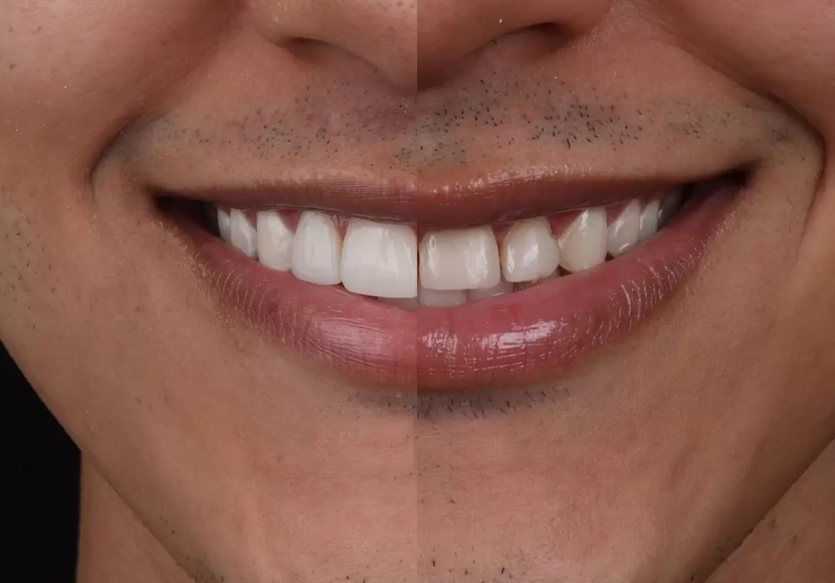
Gummy Smile Expressions: Excessive exposure of the maxillary gingiva during speech or smiling, which leaves the clinical crowns looking abnormally short, worn, or square.
Gingival Margin Asymmetry: Uneven, disharmonious gum lines across the anterior aesthetic zone, disrupting the horizontal smile arch.
2. Functional and Restorative Indications (Structural Foundation)
Subgingival Caries and Deep Fractures: Structural decay or traumatic crown fractures tracking below the bony crest, leaving insufficient supragingival tooth structure to secure an airtight restorative margin.
Inadequate Clinical Crown Height: Short clinical teeth that cannot provide enough friction or surface area to mechanically retain a permanent prosthetic crown, leading to chronic de-cementation.
How is Crown Lengthening Done? Step-by-Step Surgical Sequence
A foundational technical query analyzed by patients prior to entering clinical execution is how is crown lengthening done. The procedural execution is calibrated to the structural parameters of the defect, ranging from isolated laser soft tissue excision to full mucoperiosteal flap reflection combined with bone re-contouring:
Modality Classification | Technical Execution Blueprint | Clinical Selection Parameters |
Soft Tissue Sculpting Only (Gingivectomy) | Excess marginal gingiva is precisely trimmed and re-contoured utilizing advanced cold steel or soft tissue laser crown lengthening systems. | Selected exclusively when the underlying bone crest sits at an ideal biological distance, and the issue is pure gingival hypertrophy. |
Osseous-Supported Modification (Apically Positioned Flap + Osteotomy) | A full-thickness mucoperiosteal flap is reflected. Specialized piezoelectric (ultrasonic bone-cutting) tips harvest and trim 1-2 mm of the alveolar crest surrounding the root neck. | Mandatory when decay/fractures track below the bone crest or when the Gummy Smile phenotype is structurally bone-driven. |
The entire procedure is executed utilizing high-efficiency local anesthesia blocks, ensuring a completely painless experience, requiring approximately 30 to 45 minutes based on the volume of teeth treated. Following precise osteotomy, the tissue margins are adapted apically and secured using micro-sutures.
Comprehensive Instructions on Crown Lengthening Aftercare
The long-term success and structural stability of the repositioned tissue margins depend heavily on the patient's compliance with the after crown lengthening surgery aftercare protocol:
Immediate Post-Operative Diet Constraints: Abstain from consuming any hot foods or beverages until local anesthesia desensitization has fully resolved. Minor capillary oozing and pink-tinted saliva are fully normal for the initial 48 hours; do not spit forcefully, simply swallow naturally. Maintain a cool, soft, non-fibrous diet (yogurt, purees, chilled soups) for the initial 3-5 days.
External Cryotherapy Protocol: If surgical osteotomy (bone trimming) was incorporated, applying an external cold compress (ice pack) over the cheek for 10-minute intervals during the first day aggressively minimizes post-operative edema and swelling.
Strict Oral Hygiene Modifications: Do not apply a manual toothbrush, electric toothbrush, or dental floss directly onto the healing surgical margins for the initial 7 days. Oral decontamination of the surgical sector must be achieved via gentle rinsing with a prescribed chlorhexidine-based antimicrobial mouthwash. Normal hygiene routines must be maintained across all non-treated quadrants.
Absolute Tobacco and Nicotine Prohibition: Tobacco smoke introduces extreme heat and localized toxins that spark severe peripheral vasoconstriction, starving the healing soft tissue cells of oxygenated blood. Smoking must be strictly avoided until all sutures are professionally removed.
Frequently Asked Questions (FAQ)
How many days does recovery take after crown lengthening surgery?
The microsurgical sutures are routinely removed by our periodontist 7 to 10 days post-surgery. Once the sutures are cleared, superficial epithelial tissue healing is complete, allowing the patient to resume normal social functions. However, the internal stabilization of the alveolar bone crest and the structural maturation of the soft tissue attachment require 4 to 6 weeks to reach absolute histological finality. Consequently, fabrication of the permanent porcelain or zirconium restorations must be delayed until this biological window closes.
Does crown lengthening surgery hurt during or after the procedure?
No, crown lengthening surgery is an entirely painless procedure. Utilizing advanced, contemporary local anesthetics fully blocks all nociceptive neural paths before initiating the sequence; the patient experiences absolute zero sharp pain during tissue cutting or ultrasonic bone contouring. As the numbing agents taper off several hours later, a mild, manageable soreness or deep ache is normal, which is seamlessly resolved using basic prescribed non-steroidal anti-inflammatory drugs (NSAIDs).
Can the gum tissue grow back or relapse after a crown lengthening procedure?
When the surgical osteotomy is executed with precise milimetric accuracy to preserve the biological width relationship relative to the new bone crest, it is clinically impossible for the gum tissue to grow back or relapse. The structural transformation is permanent. However, if the patient entirely neglects daily oral plaque control post-restoration, triggering severe chronic gingivitis, inflammatory edematous gum swelling can manifest as a secondary pathological reaction.