Contentsexpand_more
- What is a Denture Sore?
- What Causes a Denture Sore?
- Symptoms of a Denture Sore
- Denture Sore Medication and Cream Treatments
- 1. Topical Gels and Creams
- 2. Antiseptic Rinses and Systemic Analgesics
- How to Heal Denture Sores: Clinical In-Office Adjustments
- Crucial Mistakes Patients Must Avoid
- Factors Influencing Denture Sore Treatment Cost
- Comparison Matrix: New Denture Sore vs. Old Denture Sore
- Frequently Asked Questions (FAQ)
- Is it normal for a brand-new denture to cause sores and pinch?
- Do denture sore creams help stick the denture in place?
- Can a chronic denture sore turn into mouth cancer?
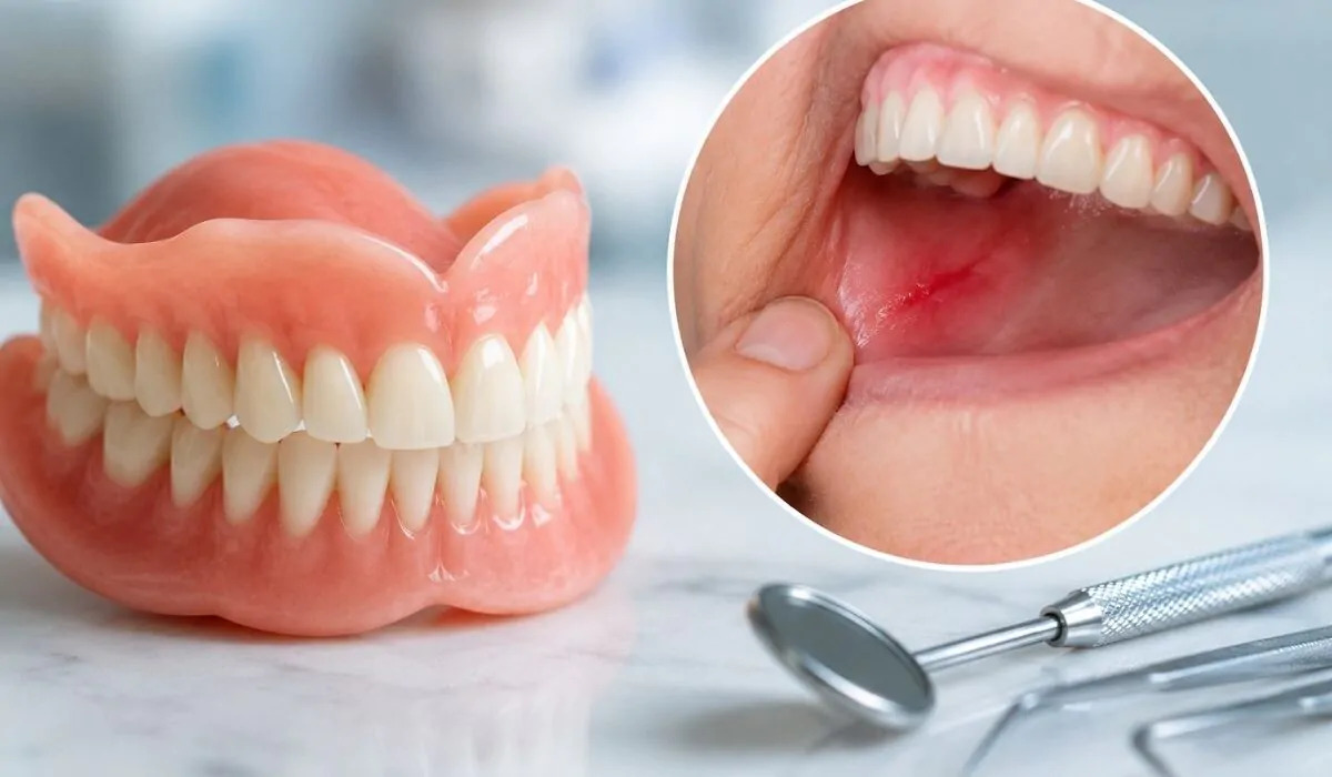
During the first several days after receiving your new complete or partial removable denture, you may experience sharp, pinching pain when chewing, speaking, or swallowing, noticing tender, whitish aphthous-like ulcers along the gingival ridge. Many patients tuck their new prostheses into a bedside drawer, anxious that their mouth will never tolerate the appliance. Much like a stiff new pair of leather shoes causing friction blisters on a heel, rigid polymethyl methacrylate acrylic resin compressing delicate, unkeratinized oral mucosa produces a clinical complication recognized as a denture sore (decubitus denture ulcer).
The oral mucosa was biologically engineered to line oral cavities, not to bear massive, continuous masticatory loads without periodontal ligament support. Unlike natural teeth that dissipate chewing forces into bone via specialized suspensory fibers, removable dentures transfer functional forces directly onto thin mucous membranes. When denture flanges are slightly overextended, an artificial tooth creates premature occlusal contact, or alveolar ridge resorption introduces instability, the underlying soft tissue is pinched.
What is a Denture Sore?
In clinical prosthodontics, the medical definition of what is a denture sore is: A localized mucosal injury characterized by epithelial necrosis, microvascular ischemia, and painful ulceration caused by mechanical pressure, shearing friction, or overextension of a removable dental prosthesis.
These lesions are not simple infectious aphthae; they are traumatic pressure sores triggered by mechanical discrepancies in the denture base. The center presents with a grayish-yellow necrotic fibrin base enclosed by an erythematous inflammatory halo, producing acute, sharp pain upon contact.
What Causes a Denture Sore?
Clinical diagnostic investigations identify several primary triggers behind denture sores:
Overextended Denture Flanges: The peripheral border of a new denture extends too deeply into the labial or lingual vestibular sulcus, slicing mucosal tissue during dynamic muscle movement.
Occlusal Disharmony and Premature Contacts: A sub-millimeter high spot on an artificial denture tooth directs disproportionate chewing forces onto one focal point of the ridge, driving the base into underlying tissue.
Alveolar Bone Spicules and Sharp Ridges: Incomplete post-extraction osseous smoothing leaves sharp cortical bony margins (knife-edge ridges) that pinch thin mucosa beneath the hard base.
Xerostomia (Severe Oral Dryness): Saliva provides a protective lubricating hydrodynamic film between acrylic and mucosa. Reduced salivary output increases interfacial friction, accelerating tissue chafing.
Residual Ridge Resorption in Older Dentures: Over time, alveolar bone resorbs while the acrylic base remains static. As gaps widen, the loose denture rocks like a seesaw during mastication, repeatedly scraping the tissues.
Wearing Dentures 24/7: Sleeping with dentures deprives mucosal tissues of essential rest and microvascular reperfusion, accelerating ulceration.
Symptoms of a Denture Sore
Typical clinical presentations associated with denture sores include:
Sharp, Needle-Like Pain on Chewing: Acute localized pinching whenever functional bite forces seat the denture base against the ridge.
Erythematous Ulcers with Whitish Slough: Discrete, painful craters with red borders and white or yellowish centers visible upon removing the appliance.
Mucosal Bleeding and Burning: Localized capillary bleeding upon cleaning the mouth, accompanied by constant stinging.
Pain Radiating to the Throat on Swallowing: Occurs when overextended lingual flanges of mandibular dentures dig into the mylohyoid or retro-mylohyoid regions.
Prosthetic Inability: Severe discomfort forcing the patient to remove the prosthesis entirely during social activities and meals.
Denture Sore Medication and Cream Treatments
While awaiting clinical adjustment, topical agents can manage pain and promote comfort:
1. Topical Gels and Creams
Anesthetic Gels: Formulations containing benzocaine or lidocaine applied 10 minutes prior to meals temporarily numb exposed sensory nerve fibers, relieving acute discomfort.
Hyaluronic Acid & Bioactive Barrier Gels: Preparations with high-molecular-weight hyaluronic acid form a protective bio-barrier that supports cell migration and accelerates tissue healing.
Corticosteroid Dental Pastes: For severe, inflamed ulcers, short-term application of triamcinolone acetonide dental paste soothes edema and speeds epithelial recovery.
2. Antiseptic Rinses and Systemic Analgesics
Warm Saline Rinses: Swishing gently with half a teaspoon of salt in warm water reduces mucosal edema and clears cellular debris.
Chlorhexidine Gluconate Rinses: Non-alcoholic antimicrobial rinses prevent opportunistic secondary bacterial colonization across the ulcer bed.
Analgesics: Over-the-counter paracetamol or ibuprofen helps control systemic inflammatory discomfort.
How to Heal Denture Sores: Clinical In-Office Adjustments
It is essential to understand: Topical medications and creams provide temporary symptomatic relief; unless the offending acrylic edge is relieved by a dentist, the sore will not resolve!
At Livera Dental Clinic, resolving a denture sore follows a precise protocol:
Pressure Point Localization: The prosthodontist coats the intaglio surface of the denture with pressure-indicating paste (PIP) or marks the intraoral ulcer with an indelible transfer pencil before reseating the appliance. The exact pressure site is identified on the acrylic base.
Micrometric Relief and Contouring: Specialized acrylic burs relieve the precise impinging borders, eliminating localized excessive thickness.
High-Gloss Polishing: The ground acrylic surface is smoothed and brought to a mirror finish using pumice and polishing wheels, ensuring no rough scratches remain.
Occlusal Balancing: Occlusal contacts are refined with articulating paper to distribute functional bite vectors evenly across the entire arch.
Denture Relining (Reline / Rebase): For older, loose dentures destabilized by bone resorption, a hard or soft laboratory reline adapts the base to current ridge anatomy, restoring stability.
Crucial Mistakes Patients Must Avoid
Do Not Attempt DIY Adjustments: Never attempt to file, shave, or cut your denture with nail files, kitchen knives, or sandpaper. DIY grinding ruins the peripheral seal, destabilizes retention, and frequently destroys the appliance beyond repair.
Wear the Denture Before Your Appointment: Keep the denture in your mouth for at least 4 to 6 hours immediately preceding your dental visit. If the denture is left out for days, the ulcer fades, making it impossible for the clinician to locate the exact contact point.
Avoid Applying Alcohol or Rubbing Agents: Never apply rubbing alcohol, perfume, or undiluted hydrogen peroxide directly onto sores; these chemicals cause secondary chemical burns and deepen the ulcer.
Factors Influencing Denture Sore Treatment Cost
Health regulations restrict stating uniform treatment fees; clinical costs depend on appliance condition:
Scope of Required Intervention: Routine spot adjustment during post-delivery warranty recalls versus complete laboratory relining or rebasing of an aged prosthesis.
Soft Silicone Relining (Permanent Cushioning): Incorporating resilient soft-silicone liners to cushion severely resorbed knife-edge ridges carries different material overhead compared to simple spot grinding.
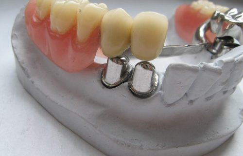
Clasp and Framework Re-adaptation: Repairing, realigning, or replacing bent cast-metal clasps in removable partial dentures.
Complete Denture Remake: If occlusal vertical dimension has collapsed and structural alignment is lost, fabricating a replacement denture may be indicated.
Comparison Matrix: New Denture Sore vs. Old Denture Sore
Parameter | New Denture Sore Spot | Old Denture Sore Spot |
Onset Profile | Days 1 to 7 post-delivery | Emerges gradually after 3 to 5 years |
Primary Etiology | Border overextension, high spot | Progressive ridge resorption, loose fit |
Denture Stability | Snug fit with localized edge pinching | Loose, rocks like a seesaw during meals |
Clinical Solution | 1 to 2 minor spot-grinding visits | Hard/soft reline or new prosthesis |
Healing Velocity | Resolves within 24–48 hours after relief | Will not resolve without base readaptation |
Medication Role | Temporary relief while mucosal drape adapts | Temporary palliative care |
Frequently Asked Questions (FAQ)
Is it normal for a brand-new denture to cause sores and pinch?
Yes. Developing minor sore spots during the first 1 to 2 weeks with a new complete or partial denture is a normal biological expectation. The mucosal tissue requires time to adapt to rigid acrylic, and initial clinical delivery cannot fully simulate every dynamic muscular movement. Dentists schedule post-insertion review appointments at 48 hours and 1 week to adjust high spots until full comfort is achieved.
Do denture sore creams help stick the denture in place?
No. Denture sore creams (such as benzocaine gels or hyaluronic acid ointments) are therapeutic medications formulated to numb pain and speed tissue healing; they contain no adhesive polymers. To stabilize a loose denture, specialized denture adhesive creams (such as Corega or Fixodent) are used. If sores are present, focus on healing the tissues and having your clinician relieve the base before relying on adhesives.
Can a chronic denture sore turn into mouth cancer?
Acute, short-lived denture sores do not cause cancer. However, ill-fitting dentures left unadjusted for years that chronically traumatize mucosal tissues can induce reactive hyperplastic overgrowths (epulis fissuratum) or chronic inflammatory cellular dysplasia. Any oral sore that fails to heal after 2 to 3 weeks despite denture removal must be evaluated by an oral surgeon to rule out neoplastic changes.