Contentsexpand_more
- What is Tooth Root Resorption?
- What Causes Tooth Root Resorption?
- Symptoms of Tooth Root Resorption
- Can Tooth Root Resorption Go Away on Its Own?
- Tooth Root Resorption Root Canal and Clinical Treatment Protocols
- 1. Management of Internal Root Resorption
- 2. Management of External Root Resorption
- Factors Influencing Treatment Cost
- Comparison Matrix: Internal vs. External Root Resorption
- Frequently Asked Questions (FAQ)
- Does a tooth with root resorption always require extraction?
- Is minor root resorption normal during orthodontic braces?
- Is the treatment for tooth root resorption painful?
During a routine dental examination or pre-prosthetic evaluation, discovering on a panoramic radiograph or cone-beam computed tomography (CBCT) scan that a clinically sound tooth possesses an hourglass thinning, an expanding hollow lumen, or a blunted root apex can be disquieting. When no dental decay is visible in the mouth, this progressive destruction occurring within the alveolar bone is clinically recognized as tooth root resorption.
While the root resorption of deciduous (baby) teeth is a physiological and natural biological mechanism enabling permanent tooth eruption, the resorption of adult permanent roots is strictly pathological. When specialized clastic cells identify the dental hard tissues as foreign substrates, progressive breakdown begins. Without prompt intervention, root resorption can lead to terminal mobility and extraction.
What is Tooth Root Resorption?
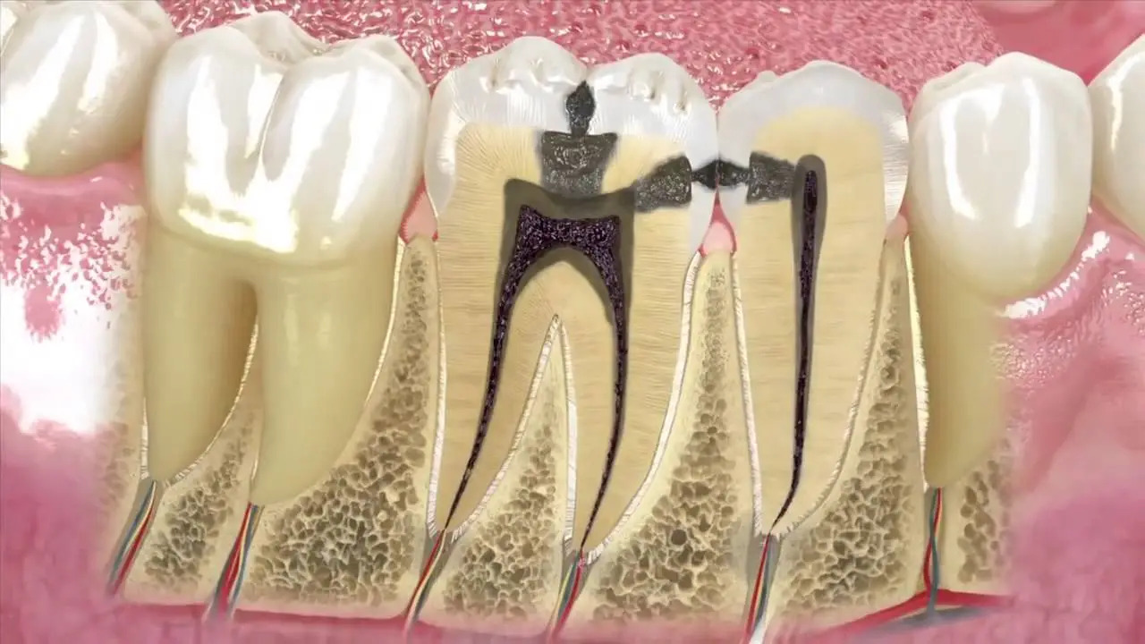
In clinical endodontics, the medical definition of what is tooth root resorption is: An inflammatory or non-inflammatory pathological process characterized by the progressive loss of dental hard tissues, namely dentin, cementum, and adjacent alveolar bone, mediated by multinucleated clastic cells (odontoclasts / osteoclasts), resulting in structural breakdown and replacement with granulation tissue.
Root resorption is classified into two primary categories based on anatomical origin:
Internal Root Resorption: Originates within the pulpal root canal space, eroding dentinal walls centrifugally from the inside out.
External Root Resorption: Initiates on the external root perimeter facing the periodontal ligament space, progressing centripetally from the outside toward the pulp.
What Causes Tooth Root Resorption?
Clinical evaluations identify several primary factors underlying what causes tooth root resorption:
Dental Physical Trauma: Remote concussive impacts, sporting accidents, or luxation injuries sustained years earlier. Damage to the protective unmineralized precementum or odontoblast layer exposes mineralized dentin, triggering clastic activation.
Excessive Orthodontic Forces: Heavy, uncalibrated mechanical vectors during fixed orthodontic bracket or clear aligner therapy can induce concentrated pressure necrosis at root apices, causing apical external resorption.
Chronic Intracanal Microbial Infection: Necrotic pulp tissue leaking bacterial toxins through apical and lateral foramina maintains chronic periapical inflammation that stimulates osteoclasts to resorb apical cementum.
Ectopic or Impacted Tooth Pressure: Impacted maxillary canines or mandibular third molars erupting in close proximity to adjacent healthy roots can exert physical pressure, inducing localized external resorption.
Complications of Internal Tooth Bleaching: Historic "walking bleach" techniques utilizing high-concentration hydrogen peroxide without a protective cervical barrier can diffuse into cervical periodontal tissues, initiating invasive cervical resorption.
Idiopathic Factors: Rare presentations where genetic predispositions or systemic conditions initiate root resorption without identifiable mechanical or microbial causes.
Symptoms of Tooth Root Resorption
Early-to-moderate root resorption is characteristically asymptomatic. When clinical markers appear, notable tooth root resorption symptoms include:
Incidental Radiographic Discovery: The vast majority of cases produce no sensations and are detected on routine periapical films or CBCT scans.
Coronal Pink Discoloration ("Pink Tooth of Mummery"): In coronal internal resorption, vascular granulation tissue undermines the thin overlying enamel, transmitting a characteristic pinkish-red hue through the crown.
Secondary Tooth Mobility: Significant structural loss along the cervical or mid-root thirds destabilizes the tooth within the alveolus.
Dull Masticatory Discomfort: Once resorption perforates the root perimeter or secondary microbial contamination occurs, localized percussion sensitivity develops.
Gingival Tissue Ingrowth: In external cervical resorption, fibrovascular tissue can breach the sulcus, presenting as an easily bleeding, hyperplastic tissue collar.
Can Tooth Root Resorption Go Away on Its Own?
Addressing whether root resorption can resolve spontaneously requires understanding dental biology:
Never Self-Limiting: Active clastic cells continue eroding dentin as long as stimulating cytokines and blood supply persist. The condition does not resolve without intervention.
Ineffective Medical Rinses: Systemic medications and topical dentifrices cannot penetrate the alveolar bone to inhibit odontoclasts.
Early Intervention Preserves the Substrate: Prompt endodontic extirpation or surgical debridement arrests the resorption, safeguarding the tooth.
Tooth Root Resorption Root Canal and Clinical Treatment Protocols
At Livera Dental Clinic, executing tooth root resorption treatment and specialized tooth root resorption root canal procedures is guided by dental operating microscopes:
1. Management of Internal Root Resorption
The therapeutic priority is total debridement of the hyperplastic pulpal tissue feeding the odontoclasts:
Under high magnification, access is established into the root canal system.
Ultrasonic irrigation utilizing warm sodium hypochlorite dissolves necrotic tissue within irregular resorptive lacunae.
Intracanal calcium hydroxide dressing is placed for several weeks to promote an alkaline environment and arrest clastic enzyme cascades.
The ballooned defect is obturated three-dimensionally using warm vertical gutta-percha compaction or bioceramic hydraulic cements (MTA / Biodentine).
2. Management of External Root Resorption
Therapeutic approach depends on lesion location:
Invasive Cervical Resorption: A localized mucoperiosteal flap is elevated, resorptive granulation tissue is curetted, the root cavitation is restored using bioceramic cements or resin-modified glass ionomers, and the flap is repositioned.
Infection-Related Apical Resorption: Standard chemo-mechanical root canal treatment eradicates intracanal microbial triggers.
Severe Structural Loss: Teeth with extensive root destruction and hopeless prognoses are slated for atraumatic extraction and implant rehabilitation.
Factors Influencing Treatment Cost
Regulatory directives prohibit publishing fixed surgical fees; clinical costs depend on case complexity. Primary variables include:
Resorption Classification and Extent: Non-perforating internal canal therapy versus complex microsurgical external cervical repairs requiring flap elevation.
Bioceramic and Regenerative Biomaterials: Volume of biocompatible Mineral Trioxide Aggregate (MTA), Biodentine, or PRF membranes required to seal defects.
Advanced Diagnostic and Operative Imaging: Utilization of localized high-resolution 3D CBCT imaging and operative dental microscopes.
Adjunctive Periodontal and Restorative Steps: Need for clinical crown lengthening, orthodontic extrusion, or post-endodontic full-coverage coronal restorations.
Comparison Matrix: Internal vs. External Root Resorption
Diagnostic Criteria | Internal Root Resorption | External Root Resorption |
Site of Origin | Pulpal canal space | Periodontal ligament surface |
Clinical Incidence | Rare | Significantly more prevalent |
Primary Etiologies | Pulpal trauma, chronic pulpitis | Orthodontic forces, trauma, impacted teeth |
Visual Hallmark | Pink coronal hue ("Pink tooth") | Often visually normal / gingival overgrowth |
Radiographic Margin | Smooth, symmetrical canal dilation | Irregular, "moth-eaten" root contours |
Primary Therapy | Root canal therapy + Bioceramics | Surgical repair / Endodontic intervention |
Frequently Asked Questions (FAQ)
Does a tooth with root resorption always require extraction?
No. An extraction is not inevitable. When diagnosed before extensive structural collapse or subgingival perforation occurs, chemo-mechanical root canal therapy paired with bioceramic obturation can arrest the defect and preserve the tooth for decades. Extractions are reserved for advanced cases with severe mobility or unrestorable cervical loss.
Is minor root resorption normal during orthodontic braces?
Slight, microscopic rounding of root apices (under 1 mm) is an accepted biological accompaniment to orthodontic tooth movement and does not compromise long-term tooth longevity. However, patients with genetic predispositions or those subjected to heavy vectors may experience pronounced shortening. Orthodontists monitor root integrity using periodic radiographic surveys.
Is the treatment for tooth root resorption painful?
No. All endodontic and microsurgical procedures are conducted under profound local anesthesia. Patients do not experience operative pain. Transient postoperative tenderness over the following 24 to 48 hours is manageable with standard over-the-counter analgesics.