Contentsexpand_more
- What is a Tooth Root Cyst?
- What Causes a Tooth Root Cyst?
- Tooth Root Cyst Symptoms
- Complications of an Upper Tooth Root Cyst
- How to Cure a Tooth Root Cyst and Modern Treatments
- 1. Non-Surgical Therapy: Tooth Root Cyst Root Canal Treatment
- 2. Surgical Therapy: Tooth Root Cyst Surgery (Apicoectomy & Enucleation)
- Comparison Matrix: Tooth Root Cyst Management Options
- Frequently Asked Questions (FAQ)
- Is tooth root cyst surgery (apicoectomy) painful or difficult?
- Does every tooth with a root cyst need to be extracted?
- Can a tooth root cyst recur after surgical removal?
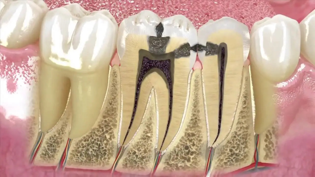
Within clinical dentistry, one of the most concerning internal pathological developments is progressive alveolar bone loss caused by untreated pulpal infections. When dental pulp tissue undergoes necrosis due to deep caries or trauma, pathogenic bacteria migrate through the apical foramen into the surrounding periapical bone. In an effort to compartmentalize and contain the bacterial invasion, the host immune response forms a fluid-filled, epithelial-lined defense capsule around the root apex. Known clinically as a periapical cyst or radicular cyst, this lesion is commonly termed a tooth root cyst.
These lesions develop asymptomatically over extended periods without causing acute pain. Patients often remain unaware of the condition until diagnostic panoramic radiographs or CBCT scans identify the radiolucency, or until the cyst perforates the cortical bone to form an active abscess. Left untreated, radicular cysts resorb significant alveolar bone volume, compromise adjacent roots, and elevate jaw fracture risks.
What is a Tooth Root Cyst?
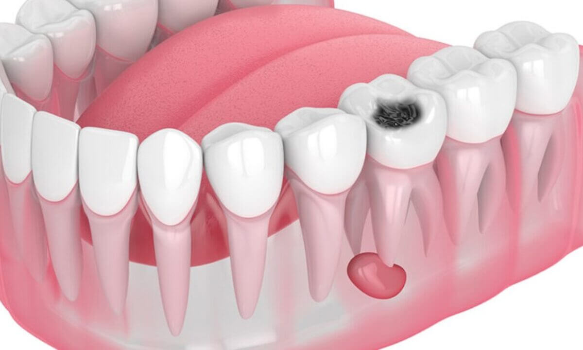
For patients seeking clinical clarity during diagnostic evaluations, the medical definition of what is a tooth root cyst is: A chronic inflammatory odontogenic cyst located at the apex of a non-vital tooth, initiated by the inflammatory stimulation of the epithelial rests of Malassez within the periodontal ligament, forming a pathological, fluid-filled cavity enclosed by a fibrous connective tissue wall and stratified squamous epithelium.
Unlike solid periapical granulomas, true cysts contain an internal cystic lumen filled with inflammatory fluid and cholesterol crystals that exert hydrostatic pressure, resorbing surrounding bone matrix.
What Causes a Tooth Root Cyst?
Evaluating what causes a tooth root cyst reveals specific microbiological and mechanical pathways:
Untreated Deep Carious Lesions: Acid-producing bacteria penetrate enamel and dentin, entering the pulp chamber and inducing pulpal necrosis.
Incomplete or Failing Endodontic Therapy: Root canals with persistent micro-leakage or uninstrumented anatomy harbor bacteria, triggering apical cyst formation years later.
Physical Dental Trauma: Blunt trauma from sports injuries, falls, or accidents severs the neurovascular bundle at the root apex, resulting in aseptic pulpal necrosis that can manifest as a chronic periapical lesion.
Severe Periodontal Disease: Retrograde infections traveling through deep periodontal pockets into the apical region.
Tooth Root Cyst Symptoms
While early-stage lesions remain asymptomatic, progressive tooth root cyst symptoms include:
Dull Pain on Mastication / Occlusal Pressure: Discomfort during chewing or when tapping (percussion) on the involved tooth.
Gingival Fistula / Parulis: A localized, pimple-like drainage tract on the gum releasing purulent exudate as the lesion vents cortical pressure.
Facial and Soft Tissue Swelling: Acute flare-ups transforming chronic cysts into active periapical abscesses with localized edema.
Intrinsic Crown Discoloration: The tooth turns dull, dark gray, or brownish, confirming loss of vitality.
Parchment Crackling (Eggshell Crackling): Severe cortical bone thinning producing a distinct crackling sensation under gentle palpation.
Tooth Mobility and Displacement: Progressive cyst expansion displacing adjacent tooth roots.
Complications of an Upper Tooth Root Cyst
An upper tooth root cyst carries unique anatomical risks due to its proximity to the midface structures:
Maxillary Sinus Perforation: Roots of maxillary molars and premolars lie in close proximity to the sinus floor. Expanding cysts can breach the Schneiderian membrane, leading to odontogenic maxillary sinusitis, unilateral nasal congestion, purulent post-nasal drip, and chronic midfacial pain.
Nasal Cavity Encroachment: Anterior maxillary cysts can extend toward the nasal floor, causing nasal breathing restriction.
How to Cure a Tooth Root Cyst and Modern Treatments
Addressing how to cure a tooth root cyst and planning tooth root cyst treatment requires clinical intervention. Cysts CANNOT be resolved with systemic antibiotics alone. Antibiotics may manage acute flare-ups, but the epithelial lining remains within the bone.
Treatment modalities include:
1. Non-Surgical Therapy: Tooth Root Cyst Root Canal Treatment
For small to moderate periapical lesions, conservative endodontic therapy serves as the primary intervention:
Root canals are chemo-mechanically debrided using operating microscopes, ultrasonic irrigation, and laser disinfection.
Long-term intracanal medicaments (such as calcium hydroxide or bioceramic pastes) are placed for 2 to 3 weeks to raise internal pH, dissolve the cystic lining, and promote osseous regeneration.
Upon radiographic confirmation of bone remodeling, canals are hermetically sealed with bioceramic sealers and gutta-percha.
2. Surgical Therapy: Tooth Root Cyst Surgery (Apicoectomy & Enucleation)
Indicated for large lesions, persistent cysts non-responsive to endodontic retreatment, or teeth with post-retained crowns:
Under local anesthesia, a mucoperiosteal flap is elevated, and a conservative osteotomy is created to expose the root apex.
The cystic capsule is enucleated with its epithelial lining intact.
The apical 3 mm of the root is resected (apicoectomy), followed by retrograde preparation and ultrasonic retro-filling with mineral trioxide aggregate (MTA) or bioceramic materials.
The bony defect is augmented with bone graft materials and barrier membranes prior to tension-free suturing.
Comparison Matrix: Tooth Root Cyst Management Options
Treatment Approach | Target Indication | Surgical Invasiveness | Preservation of Tooth | Bone Healing Window |
Therapeutic Root Canal | Small/Moderate cysts (< 1 cm) | Non-Invasive (Internal access) | 100% Retained | 3 to 6 Months |
Apicoectomy & Enucleation | Large cysts, failing root canals | Minimally Invasive (Micro-flap) | 100% Retained | 7–10 days soft tissue; 4–6 mos bone |
Tooth Extraction & Curettage | Massive cysts with >80% bone loss | Surgical Extraction | Tooth Lost (Implant planned) | 2 to 3 Months |
Frequently Asked Questions (FAQ)
Is tooth root cyst surgery (apicoectomy) painful or difficult?
No, modern apicoectomy procedures performed under local anesthesia are comfortable and pain-free. The patient feels only mild vibration or pressure. The procedure takes 20 to 30 minutes for a single root apex. Post-operative discomfort is comparable to routine dental procedures and is managed with standard prescribed anti-inflammatory medications.
Does every tooth with a root cyst need to be extracted?
No. Thanks to advancements in micro-endodontics and apical microsurgery, the majority of teeth with periapical cysts can be preserved. Extraction is reserved for cases exhibiting extensive root resorption, vertical root fractures, or complete loss of alveolar bone support where stabilization is no longer achievable.
Can a tooth root cyst recur after surgical removal?
Recurrence rates are low (below 3–5%) when complete surgical enucleation of the epithelial capsule is achieved and the root apex is sealed with retrograde bioceramics (MTA). However, if microscopic epithelial fragments remain or intra-canal micro-leakage persists, recurrence can develop over time. Performing the procedure under magnification minimizes this risk.