Contentsexpand_more
- What is Jaw Retrusion? (Mandibular and Maxillary Retrognathia)
- How to Identify Jaw Retrusion
- What Causes Jaw Retrusion?
- Do Mandibular Retrusion Exercises Work?
- At What Age Is a Jaw Retrusion Appliance Used?
- How to Fix Jaw Retrusion: Clinical Treatment Modalities
- 1. Orthopedic Growth Modification (Adolescents)
- 2. Orthodontic Camouflage (Mild-to-Moderate Adult Cases)
- 3. Jaw Retrusion Surgery (Orthognathic Surgery for Severe Cases)
- Mandibular Retrusion Before and After: Structural Transformations
- How Long Does Jaw Retrusion Treatment Take?
- Factors Influencing Jaw Retrusion Treatment Cost
- Comparison Matrix: Functional Orthopedics vs. Orthognathic Surgery
- Frequently Asked Questions (FAQ)
- Can jaw retrusion be corrected without surgery in adults?
- Does mandibular retrusion cause sleep apnea?
- Will my jaws be wired shut after jaw retrusion surgery?
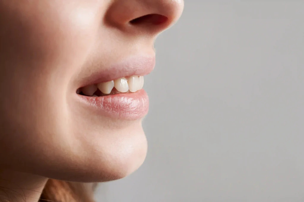
Examining your profile in lateral photographs or a three-way mirror, you may observe that your chin projection is underdeveloped, the lower lip recedes behind the nasal-mental line, and the submental tissues appear prematurely aged. Patients often assume this condition is caused strictly by protruding maxillary incisors; however, the structural defect frequently lies within the posterior position of the jawbone itself. In clinical orthodontics and orthognathic surgery, this skeletal imbalance is termed jaw retrusion (clinically diagnosed as retrognathia). Beyond compromising lateral aesthetics, jaw retrusion narrows the posterior pharyngeal airway, predisposing patients to nocturnal snoring, obstructive sleep apnea, and temporomandibular joint (TMJ) disorders.
In an aesthetically balanced facial profile, the nasal tip, lips, and chin point align harmoniously along standardized reference planes. Developmental arrest or genetic influences can disrupt this spatial equilibrium.
What is Jaw Retrusion? (Mandibular and Maxillary Retrognathia)
In dentofacial orthopedics, the scientific definition of what is jaw retrusion is: A sagittal dentofacial skeletal discrepancy characterized by the posterior displacement or hypoplasia of the mandible or maxilla relative to the cranial base and opposing jaw structures, resulting in functional malocclusion and altered facial convexity.
This condition is classified across two primary anatomical categories:
Mandibular Retrusion (Mandibular Retrognathia): The most prevalent presentation (Class II malocclusion). The upper jaw is positioned normally while the lower jaw is underdeveloped and retruded, accentuating an overjet and creating a recessed chin.
Maxillary Retrusion (Maxillary Retrognathia): Hypoplasia and posterior positioning of the upper jaw (Class III component), causing midfacial deficiency, depressed nasolabial contours, and lack of infraorbital support.
How to Identify Jaw Retrusion
Clinical diagnostic criteria used to identify jaw retrusion include:
Convex "Bird-Face" Lateral Profile: The pogonion (chin point) is positioned significantly posterior to the true vertical line, flattening the cervicomental angle and producing a double-chin appearance regardless of body weight.
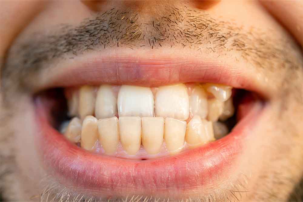
Excessive Dental Overjet: A pronounced horizontal distance between the maxillary and mandibular incisors, frequently resulting in incomplete lip competence at rest.
Airway Restriction and Sleep Apnea: Posterior displacement of the mandibular symphysis pushes the tongue base into the hypopharynx, causing nocturnal airway collapse.
TMJ Derangements and Arthralgia: The mandibular condyle is seated eccentrically within the glenoid fossa, triggering reciprocal clicking, disc displacement, and masticatory myofascial pain.
Cephalometric Radiographic Tracing: Definitive diagnosis is established through lateral cephalometric radiographs measuring standardized skeletal angles (such as ANB and SNB) against normative baselines.
What Causes Jaw Retrusion?
Diagnostic investigations identify several primary factors underlying jaw retrusion:
Genetic Heredity: Inherited craniofacial growth patterns displaying reduced mandibular ramus height or retrognathic patterns across family lineages.
Chronic Obligate Mouth Breathing: Nasal airway obstruction from hypertrophic adenoids forces open-mouth posture, directing vertical, downward-backward mandibular rotation during developmental years.
Prolonged Non-Nutritive Sucking: Thumb sucking or pacifier habits extended past age 4 restrain forward mandibular growth vectors.
Condylar Trauma and Infection: Childhood trauma to the chin or neonatal joint infections that compromise the condylar growth cartilage, arresting forward expansion.
Do Mandibular Retrusion Exercises Work?
Evaluating non-clinical techniques such as facial exercises or mewing:
No Skeletal Remodeling: Basal bone dimensions are regulated by genetics and orthopedic vector forces; voluntary facial exercises cannot lengthen the cortical mandible or advance its skeletal base.
Functional Muscular Re-education: Myofunctional therapy can assist in establishing palatal tongue posture and proper swallowing dynamics, serving as an adjunct to orthodontic therapy rather than a standalone cure.
At What Age Is a Jaw Retrusion Appliance Used?
Timing orthopedic intervention during natural growth windows is critical:
Optimal Window (Ages 9 to 14): During the circumpubertal growth spurt, the condylar cartilage responds actively to mechanical stimulation. In girls, the optimal window spans ages 10 to 12; in boys, ages 11 to 14.
Functional Orthopedic Appliances: Appliances such as Twin-Block, Herbst, Forsus, or aligners with mandibular advancement posture the lower jaw forward, stimulating remodeling within the glenoid fossa and condyle.
Limitations in Adulthood: Once the spheno-occipital synchondrosis and condylar growth plates fuse post-adolescence, orthopedic appliances can only tip teeth, not modify bone length.
How to Fix Jaw Retrusion: Clinical Treatment Modalities
At Livera Dental Clinic, how to fix jaw retrusion is categorized across three clinical approaches:
1. Orthopedic Growth Modification (Adolescents)
In growing individuals, fixed or removable functional appliances hold the mandible in an advanced position, guiding skeletal adaptation and balancing facial growth.
2. Orthodontic Camouflage (Mild-to-Moderate Adult Cases)
For mature patients seeking non-surgical options:
Fixed multibracket appliances or clear aligners retract maxillary incisors while slightly proclining mandibular incisors.
This establishes functional anterior guidance and masks the underlying skeletal discrepancy without moving the basal bone.
3. Jaw Retrusion Surgery (Orthognathic Surgery for Severe Cases)
For adults with significant skeletal discrepancies or airway collapse, surgery offers definitive skeletal repositioning:
BSSO (Bilateral Sagittal Split Osteotomy): The surgeon accesses the mandibular rami intraorally, performs controlled osteotomies, slides the lower jaw forward into ideal occlusion, and secures the bone using rigid titanium plates and screws.
Genioplasty: When occlusion is acceptable but the chin point remains recessed, an osseous genioplasty advances the inferior border of the mandibular symphysis.
Mandibular Retrusion Before and After: Structural Transformations
The functional and aesthetic improvements observed after jaw retrusion correction include:
Strong, Symmetrical Jawline: Advancing the mandibular base defines the jawline, eliminates submental soft-tissue sagging, and establishes a balanced cervical angle.
Pharyngeal Airway Expansion: Moving the jaw forward pulls the tongue base away from the posterior pharyngeal wall, resolving nocturnal airway obstruction and snoring.
Restored Masticatory Function: Correcting the Class II relationship establishes stable intercuspation and reduces anterior tooth wear.
Spontaneous Lip Competence: Eliminating excessive overjet allows the lips to close naturally at rest without mentalis muscle strain.
How Long Does Jaw Retrusion Treatment Take?
The duration of jaw retrusion treatment varies according to modality:
Functional Orthopedics in Children: Active advancement takes 9 to 12 months, followed by 12 months of fixed braces for occlusal detailing.
Adult Orthodontic Camouflage: Clear aligner or fixed appliance therapy spans 14 to 18 months.
Orthognathic Surgical Management: Comprehensive care incorporating presurgical orthodontics, the surgical osteotomy, and postsurgical detailing requires 18 to 24 months.
Factors Influencing Jaw Retrusion Treatment Cost
Health regulations prohibit advertising standardized fees; jaw retrusion costs depend on case complexity. Primary determinants include:
Clinical Modality Employed: Non-surgical functional growth modification or clear aligners versus inpatient bimaxillary orthognathic surgery.
Technological Planning Infrastructure: 3D virtual surgical planning (VSP), custom CAD/CAM surgical splints, and cone-beam CT modeling.
Operating Facility and Inpatient Care: General anesthesia, operating room infrastructure, and specialized nursing support.
Adjunctive Procedures: Combining bilateral sagittal split osteotomy with chin repositioning (genioplasty) or maxillary advancement (Le Fort I).
Comparison Matrix: Functional Orthopedics vs. Orthognathic Surgery
Feature | Functional Growth Appliances | Orthognathic Jaw Surgery |
Target Demographic | Ages 9 to 14 (Active growth) | Adults 18+ (Mature skeleton) |
Skeletal Action | Stimulates natural condylar adaptation | Controlled osteotomy and physical advancement |
Anesthesia Protocol | No anesthesia required | Inpatient general anesthesia |
Recovery Period | Immediate normal routine | 2 to 4 weeks clinical healing |
Soft-Tissue Transformation | Gradual developmental change | Immediate, significant profile correction |
Airway Impact | Early expansion of pediatric airway | Substantial, stable adult airway expansion |
Frequently Asked Questions (FAQ)
Can jaw retrusion be corrected without surgery in adults?
In mature adults, the mandibular bone cannot be stimulated to grow without surgical intervention. However, in mild-to-moderate cases where facial aesthetics are acceptable, orthodontic camouflage can retract upper teeth and adjust lower tooth inclinations to establish a functional bite without surgery. Significant skeletal discrepancies require orthognathic surgery.
Does mandibular retrusion cause sleep apnea?
Yes. When the mandible is retrognathic, the insertion point of the genioglossus muscle is displaced posteriorly, pushing the base of the tongue toward the back of the throat. This narrows the pharyngeal airway, predisposing patients to upper airway resistance syndrome and obstructive sleep apnea. Advancing the mandible expands airway volume.
Will my jaws be wired shut after jaw retrusion surgery?
No. Modern orthognathic surgery employs rigid internal fixation using biocompatible titanium mini-plates and micro-screws. This stable fixation eliminates the historic requirement of intermaxillary wire fixation, allowing patients to open their mouths, speak, and consume liquid-to-soft diets immediately following surgery.