Contentsexpand_more
- What is Oral Cancer?
- What Causes Oral Cancer?
- Oral Cancer Symptoms
- Oral Cancer Stages (TNM Staging System)
- Clinical Management of Advanced Oral Cancer
- Multimodal Oral Cancer Treatment Protocols
- Does Oral Cancer Kill You?
- Comparison Matrix: Oral Cancer Stages, Pathology & Prognosis
- Frequently Asked Questions (FAQ)
- Is every mouth sore, aphthous ulcer, or white spot cancerous?
- Does taking a biopsy cause oral cancer to spread?
- How can I lower my risk of developing oral cancer?
Intraoral ulcers, aphthous lesions, and mucosal bumps are frequently attributed to minor vitamin deficiencies, psychological stress, or accidental masticatory trauma. However, mucosal alterations that persist beyond 14 days, exhibit palpable induration, bleed spontaneously, or present with chromatic dysplasia may indicate a malignant pathological process. Representing a significant proportion of head and neck malignancies, oral cancer is curable when diagnosed at an early stage, yet carries a poor prognosis if intervention is delayed.
Developing from the malignant transformation of mucosal epithelial cells, oral carcinoma affects individuals across all age brackets, driven by tobacco, alcohol abuse, and high-risk oncogenic human papillomavirus (HPV) strains. Patients often express concerns: "What are the early signs?", "What causes oral cancer?", "Does oral cancer kill you?", and "What are the clinical survival rates?".
What is Oral Cancer?
In clinical oncology, the medical definition of what is oral cancer is: A malignant neoplastic proliferation of stratified squamous epithelial cells lining the lips, lateral and ventral tongue borders, floor of the mouth, gingiva, buccal mucosa, hard and soft palate, and retromolar trigone.
Over 90% of all oral malignancies are histopathologically classified as Oral Squamous Cell Carcinoma (OSCC). These lesions frequently arise from detectable, potentially premalignant disorders (OPMDs) such as leukoplakia (persistent white plaques) or erythroplakia (velvety red macules with high dysplasia rates).
What Causes Oral Cancer?
Addressing what causes oral cancer involves evaluating several lifestyle, viral, and chronic physical carcinogens:
Tobacco Consumption: Smoking cigarettes, cigars, pipes, and using smokeless tobacco elevates oral cancer risk by 10 to 15 times. Tobacco-specific nitrosamines induce permanent genetic mutations in tumor suppressor genes (TP53).
Excessive Alcohol Intake: Ethanol metabolizes into acetaldehyde, a direct cellular carcinogen. Alcohol dehydrates the mucosal barrier, increasing epithelial permeability to tobacco carcinogens. Concurrent tobacco and alcohol use exerts a synergistic, multiplied risk profile.
Human Papillomavirus (HPV - Strains 16 & 18): Increasingly responsible for oropharyngeal, tonsillar, and base-of-tongue carcinomas in younger, non-smoking demographics.
Chronic Mechanical Trauma: Constant mucosal laceration and chronic inflammation caused by fractured cusp margins, sharp restorations, or ill-fitting removable dentures.
Actinic Radiation (Solar UV): Direct sunlight exposure is the primary etiology for lower lip vermilion malignancies (actinic cheilitis).
Nutritional Deficiencies & Poor Oral Hygiene: Diets deficient in antioxidant micronutrients (Vitamins A, C, E, Zinc, Folate).
Oral Cancer Symptoms
Early identification hinges on recognizing clinical oral cancer symptoms:
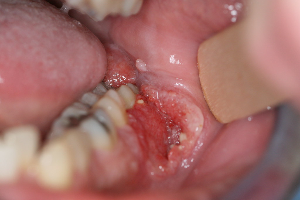
Non-Healing Mucosal Ulcers (>14 Days): Ulcerated lesions that fail to resolve within two weeks, presenting with indurated margins and central necrosis.
White (Leukoplakia) or Red (Erythroplakia) Patches: Persistent chromatic plaques that cannot be wiped away with gauze (erythroplakia carries a 70–80% malignant transformation rate).
Palpable Mass or Induration: Firm, non-tender exophytic masses or submucosal nodules within the tongue, buccal corridor, or submandibular/cervical lymph nodes.
Dysphagia, Odynophagia, and Dysarthria: Difficulty swallowing, masticating, or restricted lingual mobility accompanied by referred otalgia (ear pain).
Unexplained Dental Mobility: Sudden loosening of teeth without antecedent periodontal pocketing, caused by malignant osseous destruction.
Paresthesia / Numbness: Unexplained neurosensory loss in the lower lip, chin, or tongue due to perineural tumor invasion.
Spontaneous Hemorrhage and Fetid Halitosis: Unprovoked bleeding and necrotic tissue odors.
Oral Cancer Stages (TNM Staging System)
Clinical staging follows the TNM classification (Tumor dimensions, Regional Lymph Node metastasis, Distant Metastasis) to categorize oral cancer stages:
Stage 0 (Carcinoma in Situ): Malignant cells are confined strictly to the superficial epithelium without basement membrane penetration.
Stage 1: The primary tumor measures 2 cm or less in greatest dimension, with no regional lymph node metastasis (T1 N0 M0).
Stage 2: The tumor measures between 2 cm and 4 cm, with no lymph node involvement (T2 N0 M0).
Stage 3: The primary tumor exceeds 4 cm OR measures any size with spread to a single ipsilateral cervical lymph node measuring 3 cm or less (T3 N0 M0 or T1-T3 N1 M0).
Stage 4 (Advanced Oral Cancer): Extensive tumor invasion into cortical bone, deep extrinsic lingual musculature, skin, multiple bilateral cervical lymph nodes, or distant organs (lungs, liver, bone).
Clinical Management of Advanced Oral Cancer
Advanced oral cancer (Stages 3 and 4) represents an extensive disease state threatening functional airway, speech, and deglutition mechanics. Complications include pathological mandibular fractures, massive fixed cervical lymphadenopathy, severe facial disfigurement, and cachexia. Treatment requires a multidisciplinary tumor board combining maxillofacial oncologic surgeons, reconstructive microsurgeons, medical oncologists, and radiation oncologists.
Multimodal Oral Cancer Treatment Protocols
Modern oncologic management dictates oral cancer treatment based on disease stage and anatomical location:
Surgical Resection and Neck Dissection: The foundational modality for resectable tumors. The lesion is excised with 1.0 to 1.5 cm microscopic clear surgical margins. Selective or comprehensive neck dissection removes regional lymph node basins. Complex defects are reconstructed using microvascular free tissue transfer (e.g., vascularized fibula free flaps or radial forearm flaps).
Radiation Therapy (External Beam Radiotherapy): High-energy ionizing radiation (IMRT) eradicates residual microscopic neoplastic cells post-surgery or serves as definitive therapy in non-surgical candidates.
Chemotherapy and Targeted Immunotherapy: Systemic cytotoxic agents (Cisplatin, 5-FU) and checkpoint inhibitors (PD-1 blockers like Pembrolizumab) administered concurrently with radiation (chemoradiotherapy) in advanced-stage presentations.
Does Oral Cancer Kill You?
Addressing does oral cancer kill you, oncology outcomes demonstrate: Oral cancer is not fatal when detected at an early localized stage; however, delayed diagnosis carries high mortality.
Early Detection (Stages 1 & 2): 5-year relative survival rates range between 85% and 90%.
Advanced Stages (Stages 3 & 4): Once regional nodal metastasis or distant dissemination occurs, 5-year survival rates drop to 40% to 50%.
Undergoing routine biannual dental examinations that include systematic mucosal soft tissue screening is essential for early diagnosis.
Comparison Matrix: Oral Cancer Stages, Pathology & Prognosis
Clinical Stage | Primary Tumor Size (T) | Nodal Metastasis (N) | Distant Spread (M) | 5-Year Relative Survival |
Stage 1 | ≤ 2 cm in diameter | No nodal involvement (N0) | Absent (M0) | 85% – 90% |
Stage 2 | 2 cm to 4 cm | No nodal involvement (N0) | Absent (M0) | 70% – 80% |
Stage 3 | > 4 cm or any T with | Single ipsilateral node ≤3 cm (N1) | Absent (M0) | 50% – 60% |
Stage 4 (Advanced) | Infiltrating bone, skin, deep muscle | Multiple/Bilateral nodes (N2/N3) | Present or Absent (M0/M1) | 30% – 45% |
Frequently Asked Questions (FAQ)
Is every mouth sore, aphthous ulcer, or white spot cancerous?
No. Over 95% of common intraoral ulcers are benign aphthous stomatitis, mechanical traumatic ulcers, or viral lesions caused by stress, nutritional deficiencies, or accidental biting. Benign ulcers resolve spontaneously within 7 to 10 days. However, if an ulcer, red plaque, or white patch persists longer than 14 days, exhibits firm margins, and does not heal, an immediate clinical examination and incisional biopsy by an oral surgeon are mandatory.
Does taking a biopsy cause oral cancer to spread?
No. This is a hazardous medical myth. An incisional or punch biopsy removing a small specimen of tissue under local anesthesia is the definitive, gold-standard diagnostic tool required to confirm malignancy, determine histopathologic grading, and guide life-saving treatment. Biopsies performed under sterile clinical protocols do not disseminate neoplastic cells; rather, they prevent catastrophic delays in therapy.
How can I lower my risk of developing oral cancer?
Primary preventive measures include: Completely abstaining from all forms of tobacco, moderating alcohol consumption, maintaining a diet rich in antioxidant cruciferous vegetables and fruits, wearing UV-blocking lip balms, receiving the HPV vaccine, replacing ill-fitting dentures or smoothing jagged teeth, and scheduling routine dental check-ups every 6 months for professional soft tissue screening.