Contentsexpand_more
- What is Congenital Missing Teeth?
- What Causes Congenital Missing Teeth?
- 1. Genetic Mutations and Hereditary Inheritance
- 2. Intrautero and Environmental Disruptions
- Congenital Missing Lateral Incisor: Functional and Aesthetic Repercussions
- How to Identify Symptoms of Congenital Tooth Agenesis?
- How is Hypodontia Treated?
- 1. Advanced Orthodontic Alignment
- 2. Advanced Dental Implant Interventions
- 3. Resin-Bonded Maryland Bridges and Cosmetic Contouring
- Frequently Asked Questions (FAQ)
- If a primary baby tooth is congenitally missing, will the permanent replacement tooth still erupt?
- How long can a retained baby tooth remain functional in the mouth?
- Is dental implant surgery mandatory to correct congenital missing teeth?
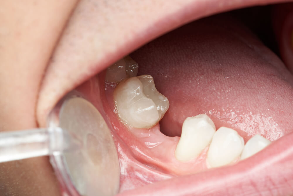
During pediatric development, the exfoliation of deciduous (baby) teeth followed by the functional eruption of permanent adult teeth represents a highly anticipated biological milestone. However, in certain clinical instances, specific permanent tooth buds completely fail to manifest within the jawbone. Discovered either through direct visual inspection in the mirror or during a routine digital panoramic radiographic screening, this developmental anomaly is clinically defined as tooth agenesis or hypodontia, and commonly referred to as congenital missing teeth.
Leaving these spaces untreated compromises more than just smile symmetry; it leads to severe masticatory inefficiency, forces adjacent teeth to tip into the edentulous space, induces localized alveolar bone resorption, and provokes speech articulation errors (phonetic variations). Fortunately, with contemporary digital orthodontics and implantology, this heritable anomaly can be fully corrected through highly synchronized clinical protocols aligned with the patient's growth phase. This extensive guide evaluates the underlying genetic etiologies and advanced structural treatments for missing tooth buds.
What is Congenital Missing Teeth?
Human tooth formation initiates deep within the embryonic tissue via specialized building structures known as tooth germs. Congenital missing teeth describe a structural anomaly where these localized buds fail to differentiate during embryonic or early pediatric development phases due to genetic signaling failures or environmental cell disruption. In clinical parameters, tooth agenesis is classified according to the volume of missing teeth:
Hypodontia: The congenital absence of 1 to 5 permanent teeth, excluding the third molars (wisdom teeth). This represents the most widespread presentation managed at our department.
Oligodontia: The genetic failure of 6 or more permanent tooth germs to form across the arches. This presentation is routinely associated with complex systemic ectodermal syndromes.
Anodontia: An exceptionally rare genetic anomaly characterized by the absolute absence of all primary and permanent dentition.
The teeth exhibiting the highest statistical frequency of congenital absence include the third molars, the maxillary permanent lateral incisors, and the mandibular second premolars.
What Causes Congenital Missing Teeth?
The structural failure of a tooth germ to initiate within the alveolar ridge is a multifactorial process. Based on clinical screenings, congenital missing teeth reasons are split into two primary biological fields:
1. Genetic Mutations and Hereditary Inheritance
The leading answer to what causes congenital missing teeth is hereditary inheritance. Mutations within specific homeobox genes that coordinate early tooth bud patterning (including MSX1, PAX9, AXIN2, and EDA) halt the development of the tooth germ at a molecular level. If a primary biological parent presents a history of missing permanent teeth, the statistical likelihood of manifestation in the offspring escalates significantly. Furthermore, tooth agenesis operates as a core symptom in several systemic syndromic conditions, such as Ectodermal Dysplasia, Down Syndrome, and Cleft Lip and Palate.
2. Intrautero and Environmental Disruptions
Severe maternal infections during gestation, high-dose localized radiation exposure, unregulated pharmacological intake, acute nutritional deficits (specifically disruptions in Vitamin D and Calcium absorption), or severe systemic pediatric illnesses during early mineralization phases can permanently damage delicate tooth germs, disrupting their development.
Congenital Missing Lateral Incisor: Functional and Aesthetic Repercussions
The clinical presentation that exhibits the most immediate, severe impact on anterior smile design is the congenital missing lateral incisor anomaly. The lateral incisors are the critical aesthetic teeth positioned immediately adjacent to the central front incisors.
When these buds are genetically absent, the sharp permanent maxillary canines (eyeteeth) naturally migrate mesially into the vacant lateral spaces. Because canines possess a distinctively yellowish hue, sharp cusps, and bulky root bulges, their anterior migration creates a highly asymmetrical, unharmonious, and structurally aggressive smile line. Furthermore, this migration provokes significant dental midline shifts and leaves expansive gaps (diastemas) between the remaining front teeth, inducing severe psychological self-esteem issues in young and adult patients.
How to Identify Symptoms of Congenital Tooth Agenesis?
The primary indicators of structural agenesis become evident as a child transitions into permanent dentition phases:
Prolonged Retention of Primary Teeth: Physiologically, an erupting permanent tooth dissolves the roots of the primary tooth directly above it, causing it to shed. If no underlying permanent bud exists, the deciduous tooth remains structurally stable and can linger in the arch well into a patient's 20s or 30s.
Asymmetrical Eruption Cycles: If a specific tooth fully erupts on the right side of the arch, but its exact anatomical counterpart on the left side fails to surface after several months, radiographic evaluation is required.
Definitive Panoramic Radiographic Findings: Absolute diagnosis is established solely via digital imaging. If the panoramic radiograph confirms zero root structure or internal tooth germ shadows within the alveolar bone matrix, structural agenesis is validated.
How is Hypodontia Treated?
Rehabilitating genetic tooth agenesis successfully cannot be achieved through an isolated dental procedure. Hypodontia treatment mandates a highly synchronized, multi-disciplinary approach uniting Orthodontists, Maxillofacial Surgeons, and Prosthodontists. The final treatment architecture is strictly dictated by the patient's chronological age and skeletal maturity:
1. Advanced Orthodontic Alignment
As the primary phase of structural therapy, the orthodontist executes one of two strategic diagnostic pathways based on facial profile analytics and skeletal space ratios:
Space Closure Strategy: If the missing tooth volume is highly localized and the skeletal profile permits, braces are utilized to guide the posterior teeth forward, fully closing the gaps. The transitioned canines are then re-shaped via cosmetic contouring to mimic natural lateral incisors.
Space Opening and Preservation Strategy: If a permanent dental implant is indicated, orthodontic forces are deployed to correctly realign the adjacent roots to their physiological angles, generating a precise, millimetric space configuration required to receive a titanium implant.
2. Advanced Dental Implant Interventions
Once craniofacial skeletal growth is fully completed (typically after age 18 in females and 20 in males), inserting a premium titanium dental implant into the orthodontically prepared space stands as the gold standard solution. The implant acts as a prosthetic root, preserving the surrounding alveolar ridge from disuse resorption. It is capped with a custom CAD/CAM zirconia or porcelain crown, delivering immaculate structural anatomy that seamlessly integrates with the surrounding natural teeth.
3. Resin-Bonded Maryland Bridges and Cosmetic Contouring
For adolescent patients who have completed active orthodontic alignment but have not yet reached the age threshold mandatory for safe dental implant placement, the created spaces must be preserved. Utilizing Maryland Bridges where a custom ceramic tooth is bonded directly to the internal lingual surfaces of adjacent teeth using micro-wings without altering healthy enamel provides an exceptional, highly aesthetic interim restoration that preserves space until surgical maturity is achieved.
Frequently Asked Questions (FAQ)
If a primary baby tooth is congenitally missing, will the permanent replacement tooth still erupt?
If a primary deciduous tooth is structurally absent from birth, the statistical likelihood that its permanent counterpart is also genetically missing exceeds 90%. However, definitive structural confirmation can only be verified via a panoramic radiographic analysis.
How long can a retained baby tooth remain functional in the mouth?
In the absolute absence of an underlying permanent tooth germ, a retained baby tooth can successfully serve masticatory functions well into a patient’s 30s or 40s, provided the patient maintains excellent oral hygiene and the root structure remains free of inflammatory resorption. These teeth must be preserved as long as they remain stable.
Is dental implant surgery mandatory to correct congenital missing teeth?
No, it is not mandatory. However, it represents the most conservative, highly reliable modern therapy because it eliminates the requirement to cut down adjacent healthy teeth to receive a bridge while actively preventing localized jaw bone resorption. For patients rejecting surgery, porcelain laminates, bonding, or classic bridge prosthetics remain viable options.