A Panoramic Dental X Ray is known for displaying the entire oral and jaw structure in a single frame, making it one of the most commonly used imaging techniques in modern dentistry. This method offers a fast overview of the teeth, jawbones, and surrounding tissues, which is why it has been an area of interest for both specialists and patients. Although sometimes referred to as dental panoramic imaging or jaw panorama, the most recognized term remains Panoramic Dental X Ray. Its ability to provide broad, practical, and interpretable data distinguishes it from other imaging techniques.

Panoramic Dental X Ray and the General View of the Oral Structure
A Panoramic Dental X Ray captures all structures in the mouth in one scan and is particularly useful during initial evaluations. Rather than examining a single tooth, its purpose is to present a panoramic perspective that connects each structure with the others, making the general anatomy easier to understand.
With the rise of digital systems, panoramic scans now offer improved resolution, contrast, and detail. Modern devices can produce clearer results with lower radiation levels. The generated image can be processed through various analysis software, allowing more refined insights into jaw symmetry, bone density, and tooth positioning. This contributes to its growing use in clinical research and dental education.
Why Is a Panoramic Dental X Ray Used?
Many people wonder why the Panoramic Dental X Ray has become such a standard tool. Its popularity stems from being quick, comprehensive, and adaptable. Conditions that cannot be easily noticed during a basic clinical examination can become visible through panoramic imaging. Tooth alignment, bone shape, and density variations can all be assessed from this single frame.
This method is particularly advantageous for examining impacted teeth. Deeply positioned tooth roots, jaw joint structure, or hidden bone formations become more interpretable through panoramic scans. It is also frequently used in orthodontic planning to understand skeletal relationships before treatment begins. From jaw surgery to periodontal assessments, panoramic imaging serves as a supportive diagnostic tool.
How Is a Panoramic Dental X Ray Taken?
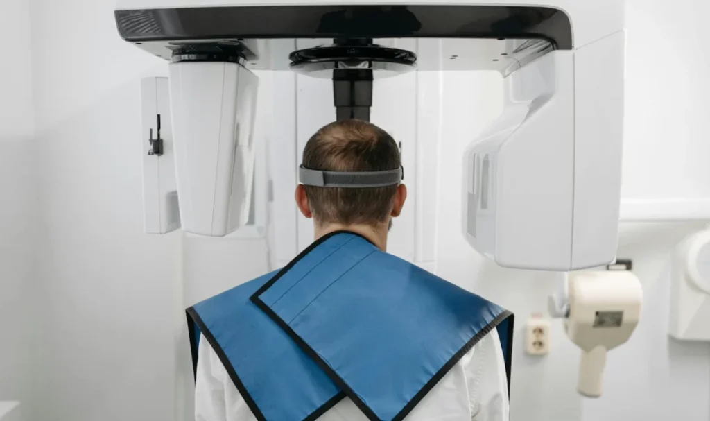
A Panoramic Dental X Ray is a quick, simple procedure that usually takes only a few minutes. The individual stands in the machine and positions their chin using stabilizing supports. This stabilization is crucial because the imaging arm rotates around the head and requires minimal movement to produce a clear result.
During the scan, the person must remain still. Digital systems display the image immediately after the scan, allowing specialists to zoom, measure, and adjust contrast. Because no extensive preparation is required, the procedure can easily fit into daily clinic routines.
When Is a Panoramic Dental X Ray Preferred?
Although it is not performed on every patient, a Panoramic Dental X Ray is preferred when a general assessment of the entire jaw is necessary. It supports clinical examination when anomalies in tooth development or jaw structure are suspected. It also helps monitor structural changes in the oral cavity over time.
In orthodontic planning, evaluating the relationship between tooth alignment and jaw structure is essential. Panoramic imaging provides the broad perspective needed at the beginning of treatment. It is also valuable in examining impacted teeth, identifying cyst-like formations, and planning dental implants. While it may not offer fine detail for small lesions, it is ideal for wide scale evaluations.

Advantages of a Panoramic Dental X Ray
A Panoramic Dental X Ray allows researchers and clinicians to compare various oral structures simultaneously, saving time during the diagnostic process. Tooth positioning, jawbone form, and joint features can all be evaluated at a glance.
Digitized panoramic scans can be analyzed rapidly. Regions of interest can be highlighted, tissue density differences can be examined, and multiple measurements can be performed. Population based research studies also benefit from panoramic imaging to investigate jaw asymmetry, dental development, and bone density patterns.
Limitations of a Panoramic Dental X Ray
Every imaging method has limitations, and the Panoramic Dental X Ray is no exception. Because it captures a wide area, it may not always provide high detail for small structures. Some findings may require more sensitive imaging techniques.
If the patient moves during the scan, the image may become blurred. Additionally, the wide angle format may cause overlapping of certain structures. Even with these limitations, panoramic imaging remains a valuable tool for broad clinical and academic observation.
Analytical Use of Panoramic Dental X Rays
A Panoramic Dental X Ray does more than image the oral structures. It creates an analytical foundation by enabling measurements of tooth positions, jaw proportions, and density variations. These measurements help interpret jaw symmetry, tooth arrangement, and general oral development from a broader viewpoint.
Researchers also use panoramic scans to compare differences across age groups, bone structures, and dental development periods. In clinical settings, panoramic imaging serves as the starting framework for understanding the overall situation before focusing on more specific details.
The Role of Panoramic Dental X Rays in Clinical Studies
Panoramic Dental X Rays are valuable in studies with large sample groups, such as population based jaw structure research. Many academic investigations rely on panoramic images to assess tooth orientation, age related bone changes, and full arch anatomical variations.
In educational environments, having the entire oral structure in a single frame helps students gain a conceptual understanding of dental anatomy. They can observe jaw joints, sinus outlines, impacted teeth, and bone form more clearly.
A Panoramic Dental X Ray offers a wide angle view of the oral and jaw structures and plays an essential role in both clinical and academic fields. It helps visualize how teeth and bones relate to each other and provides a starting point for broader assessment. Although it does not always offer detailed information about small regions, it excels at presenting an overview that guides further analysis.
With the continuous improvement of digital imaging technologies, panoramic scans are becoming clearer and more precise. This development enhances their usefulness in clinical observations and research based evaluations. Thanks to the broad perspective they offer, panoramic images will likely remain an important tool for examining oral and maxillofacial structures for many years.


